Pes cavus is a foot deformity with an unusually high arch, and it is sometimes the first sign of a nerve or muscle condition. Its causes, symptoms, assessment and treatment options.
On this page 8 sections
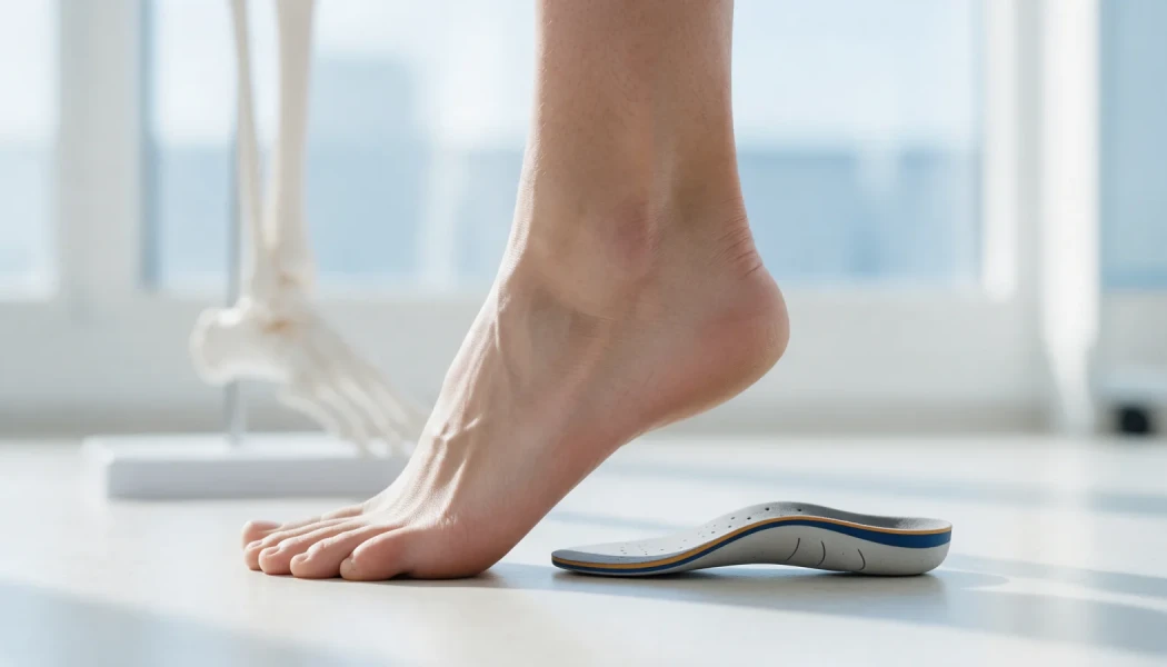
Pes cavus is a foot shape in which the inner arch is higher than normal and does not flatten when you stand; it is often combined with a heel that tilts inwards, an alignment known as "cavovarus". A mild high arch that causes no symptoms may simply be a person's natural foot type. However, when pes cavus is marked, getting worse or present in only one foot, it is advisable to look for an underlying nerve or muscle condition. Treatment usually starts with suitable insoles, footwear and exercises; for marked deformity and persistent symptoms, soft-tissue and bone-correction surgery may be considered.
This article explains how a high-arched foot is recognised, what can cause it, the problems it may lead to and the treatment options considered for children and adults.
What is pes cavus and how is it recognised?
In a normal foot the inner arch flattens slightly under load, spreading body weight evenly between the heel, the outer edge of the foot and the forefoot. In pes cavus the arch is high and the foot contacts the ground mainly at the heel and the forefoot. Pressure is therefore concentrated on a small area, particularly under the heads of the metatarsal bones and along the outer edge of the foot.
Clues that suggest a high arch include:
- A footprint with a very narrow middle section, or none at all
- Being able to see the inner side of the heel when looking at the foot from the front, a sign that the heel tilts inwards
- Clawing of the toes
- Shoes wearing down quickly along the outer edge
The deformity may be flexible or may stiffen over time. In a flexible deformity the joints can be corrected by hand; in a rigid one they cannot. This distinction is central to treatment planning.
Causes of pes cavus: why investigation matters
When the balance between the muscles that move the foot is disturbed, the foot gradually takes on a high-arched shape. For example, if the muscles that lift the foot and turn it outwards become weak while the muscle that pushes the first metatarsal down stays strong, the forefoot bends downwards, the heel tilts in and the toes claw. This is why a significant proportion of people with marked pes cavus have an underlying neurological condition.
| Group of causes | Examples | Features that stand out |
|---|---|---|
| Inherited nerve conditions | Charcot-Marie-Tooth disease (hereditary motor and sensory neuropathy) | Usually both feet, similar feet in the family, thinning of the lower legs, later weakness of the hand muscles |
| Spinal cord and central nervous system | Tethered spinal cord, cysts or growths within the spinal cord, cerebral palsy, spina bifida | One-sided or progressive, signs on the lower back, bladder or bowel problems |
| Congenital or after earlier treatment | Residual deformity after clubfoot treatment | History of treatment in childhood |
| After injury | Malunion after foot or leg fractures, muscle contracture after compartment syndrome | One-sided, starting after a specific injury |
| Familial or no cause found | High arch with no underlying condition identified | Usually mild, on both sides and not progressing |
Because children treated for clubfoot can develop a high arch in later years, follow-up matters; you can read more in our clubfoot treatment guide.
What problems can pes cavus cause?
Because a high-arched foot carries load over a smaller area, it can lead to various problems over time:
- Pain and calluses under the metatarsal heads and along the outer edge of the foot
- Repeated ankle sprains and a feeling of instability
- Claw toes, with sores on top of the toes where shoes rub
- Difficulty finding comfortable shoes
- Heel pain and tightness in the sole of the foot
- Pain in the tendons on the outer side of the foot, and stress fractures of the fifth metatarsal
- Arthritis of the ankle and foot joints in the long term
When pes cavus is caused by a nerve condition, it may also come with the toes catching on the ground when walking, frequent falls and reduced sensation in the feet.
Assessment: examination and tests
Assessment begins with a detailed history and examination. The doctor will ask how long the symptoms have been present, whether they are getting worse and whether anyone in the family has similar feet or a nerve condition. The examination covers the appearance of the foot standing and sitting, joint flexibility, muscle strength, reflexes, sensation, the spine and the hands.
An important part of the examination is the Coleman block test. You stand with the outer edge of the foot and the heel on a thin block, while the first metatarsal is allowed to drop off its edge. If the inward tilt of the heel corrects, the hindfoot is flexible and the deformity is mainly driven by the forefoot; if it does not, the hindfoot has stiffened. If surgery is needed, this helps to decide which part of the foot should be corrected.
Tests are planned for the individual and may include:
- Standing (weight-bearing) X-rays of the foot and ankle
- Special X-ray views showing heel alignment where needed
- Nerve conduction studies and a neurology opinion if a nerve condition is suspected
- Genetic testing in appropriate cases
- An MRI of the spine for a one-sided or rapidly progressing deformity
Pes cavus treatment without surgery
For many people with mild symptoms or a flexible deformity, treatment begins without surgery. The aims are to spread pressure more evenly across the sole, support the ankle and maintain muscle balance:
- Custom insoles: cushioned insoles that spread pressure and, where needed, offload the area under the first metatarsal head can help reduce pain and callus formation.
- Suitable footwear: shoes with a wide, deep toe box and a cushioned sole; if the ankle feels unstable, styles that support the ankle may help.
- Orthoses: ankle braces or ankle-foot orthoses may be used for people with recurrent sprains or foot drop.
- Exercise and physiotherapy: stretching of the calf and sole, strengthening of the muscles that turn the foot outwards, and balance training.
- Skin care: regular care of calluses and, for people with reduced sensation, checking the feet every day.
In children whose pes cavus is caused by a nerve condition, the deformity can increase with growth, so regular follow-up is important.
Surgery for pes cavus: when and which procedures?
Surgical options are considered when pain, recurrent sprains, a deformity that makes shoes difficult to wear, a risk of skin breakdown or progression continue despite non-surgical treatment. The aim of pes cavus surgery is not simply to lower the arch but to restore a balanced, flat-footed stance and muscle balance, so several procedures are often combined:
- Soft-tissue releases: releasing the tight band of tissue in the sole of the foot (the plantar fascia)
- Tendon transfers: moving the tendon of a strong muscle so that it takes over the role of a weak one and rebalances the foot
- Osteotomies: controlled cuts to realign the first metatarsal or the heel bone
- Toe corrections: straightening claw toes with tendon and joint procedures
- Joint fusion (arthrodesis): for severe, rigid deformities, usually in adults, fusing certain foot joints in a corrected position
In children, joint-preserving methods are preferred wherever possible, and remaining growth is taken into account. For some patients who have had previous surgery, have fragile skin or soft tissues, or have a very severe deformity, gradual correction over days to weeks with a circular external fixator (Ilizarov frame) can be an option. For the approach to foot and leg deformities in adults, see our pages on adult leg deformity correction and correction of leg, knee and ankle deformities; for children, see childhood lower limb deformities.
After surgery a cast or boot is usually worn for several weeks, weight-bearing is increased gradually and physiotherapy follows. The time needed to return to daily activities and sport depends on the procedures performed and varies from person to person.
When should you see a doctor?
An orthopaedic assessment is advisable if you notice:
- A high arch or a change in foot shape on one side only
- An arch that becomes higher over time, with clawing of the toes, in a child or an adult
- Frequent ankle sprains, toes catching when walking or frequent falls
- Thinning, weakness or reduced sensation in the legs or hands
- A family history of similar feet or an inherited nerve condition
- Pain and calluses that persist despite insoles and suitable shoes
- Any change in bladder or bowel control alongside the foot changes
Prof. Dr. Halil İbrahim Balcı worked as a research fellow in deformity surgery and Ilizarov techniques at the Paley Institute in the United States. In pes cavus, the treatment plan is based on the examination and test results and, where needed, is coordinated with other specialties such as neurology.
Key points
- Pes cavus is a foot deformity with an unusually high arch, often with the heel tilted inwards.
- Marked, progressive or one-sided pes cavus should prompt a search for an underlying nerve or muscle condition.
- The Coleman block test and weight-bearing X-rays help show how flexible the deformity is and where it originates.
- Treatment usually starts with insoles, footwear, orthoses and exercise.
- For persistent symptoms, individual surgery combining soft-tissue, tendon and bone procedures may be planned.
Frequently Asked Questions
In flat feet (pes planus) the inner arch collapses noticeably when standing and most of the sole touches the ground. In pes cavus the arch is high, the foot rests mainly on the heel and forefoot, and pressure is concentrated on a small area. The two conditions have different causes, symptoms and treatments; examination and, if needed, weight-bearing X-rays make the distinction clear.
Pes cavus is not always hereditary, but in some families similar feet appear over several generations. In particular, one of the common causes of progressive pes cavus affecting both feet is Charcot-Marie-Tooth disease, an inherited nerve condition. If there are similar feet in the family, or thinning or weakness of the legs, a neurology assessment and, where appropriate, genetic testing may be recommended.
For pes cavus, cushioned insoles made for the individual foot, which spread pressure over a wider area of the sole, are generally preferred. In some cases extra modifications are added to offload the area under the first metatarsal head or to counter the inward tilt of the heel. Off-the-shelf insoles may not suit every foot, so it is sensible to have them prescribed after an examination.
Recovery time depends on the type and number of procedures performed. When bone procedures are carried out, a cast or boot is usually worn for several weeks, with limited weight-bearing during this time. Gradual weight-bearing and physiotherapy follow. A return to everyday walking often takes a few months, while a return to sport may take longer. The process differs from person to person.
Many people with a mild high arch that causes no symptoms can take part in sport. However, if you have recurrent ankle sprains, pain along the outer edge of the foot or stress fractures, suitable shoes, insoles and balance and strengthening work become important. If symptoms increase with running or jumping sports, an examination is recommended to identify the cause and appropriate measures.
This article is for general information only and does not replace a medical examination, diagnosis or treatment. Treatment options and outcomes vary from person to person.
Related treatment area Childhood Lower Limb Deformities: Bow Legs, Knock Knees and In-Toeing