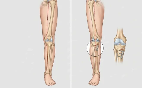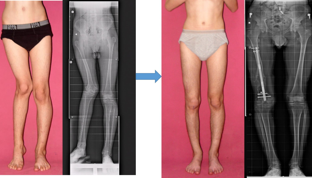
Leg deformity correction in adults straightens bow legs (genu varum) or knock knees (genu valgum) by cutting the bone in a controlled way and fixing it again at the correct angle, an operation known as an osteotomy. The aim is not only to change how the legs look but, more importantly, to make body weight pass evenly through the knee, protect the overloaded compartment and relieve symptoms. The most common procedures are a high tibial osteotomy (HTO) at the top of the shin bone and a distal femoral osteotomy (DFO) at the lower end of the thigh bone; the correction may be held with a plate, a nail or an external fixator.
This page covers leg deformities in people whose growth is complete: their causes, their effect on the knee, how they are measured, the treatment options, recovery and the risks. Physiological and disease-related bowing in children, and treatments that guide growth, are explained on our page on childhood lower limb deformities.
What are bow legs and knock knees in adults?
When you stand, a line drawn from the centre of the hip to the centre of the ankle is called the mechanical axis. In normal alignment, this line passes close to the middle of the knee. In bow legs it shifts towards the inner side of the knee, and in knock knees towards the outer side. Mild variation between individuals is normal; mild bowing in particular is very common and does not always need treatment.
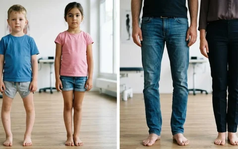
With bow legs, a gap remains between the knees when the ankles are together; with knock knees, a gap remains between the ankles when the knees are together. Appearance can be misleading, though: the shape of the calf muscles, the distribution of soft tissue and rotational differences in the bones can make a leg look more or less curved than it really is. Assessment is therefore always based on X-ray measurements.
What causes leg deformity in adults?
- Constitutional alignment: a natural alignment that has been present since childhood and often runs in families.
- Childhood deformity that never corrected or has recurred: for example after rickets, Blount disease or growth plate damage.
- Malunion after a fracture: a fracture around the knee or in a long bone that has healed at an angle.
- Knee osteoarthritis: as cartilage is lost, the inner compartment collapses and bowing increases; collapse of the outer compartment increases knock knees.
- Previous meniscus removal: after a meniscectomy, load and wear in that compartment can increase.
- Metabolic bone disease and skeletal dysplasias: conditions such as osteomalacia (softening of the bones), vitamin D-resistant rickets or achondroplasia.
How does deformity affect the knee, and which symptoms matter?
Even in normal alignment, the inner compartment of the knee carries the larger share of body weight. With bow legs this load becomes concentrated even further on the inner side; with knock knees, the outer compartment is overloaded. Over the years, cartilage and meniscus in the overloaded compartment can wear more quickly. As they wear, the deformity increases, and as the deformity increases so does the load, creating a vicious circle. Correcting marked deformity at the right time may therefore help, in suitable patients, to slow the progression of arthritis and postpone the need for a knee replacement, although this effect is not the same for everyone.
Symptoms that matter include:
- Pain on the inner side of the knee (with bow legs) or the outer side (with knock knees) that builds up with walking and towards the end of the day
- Difficulty standing or walking for long periods
- A feeling that the knee shifts sideways when you walk
- A deformity that has clearly increased over the years, or is more obvious in one leg
- With knock knees, kneecap instability or pain at the front of the knee
- Ankle or hip complaints caused by a compensating gait
Assessment: measurement and imaging
In leg deformity surgery, the outcome depends to a large extent on accurate measurement and planning. Assessment includes:
- Clinical examination: the degree of deformity, ligament laxity, range of movement, bone rotation and leg lengths are assessed.
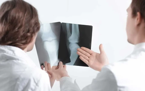
- Standing full-length leg X-ray: shows where the mechanical axis passes through the knee and allows the angles of the thigh and shin bones to be measured. This reveals whether the deformity comes from the femur, the tibia or both.
- CT scanning: used if a rotational deformity is suspected as well.
- MRI: shows the condition of the cartilage, menisci and ligaments, and helps to establish which compartment is still healthy.
Where the deformity comes from determines where it should be corrected. For example, correcting a knock knee that arises from the thigh bone by operating on the shin bone can leave the knee joint line tilted. The general principles of deformity analysis, including the centre of rotation of angulation (CORA), are explained on our deformity surgery page.
Treatment options: non-surgical care, osteotomy and joint replacement
Weight management, exercises to strengthen the thigh muscles, physiotherapy, suitable footwear and insoles and, where needed, an unloader knee brace can ease symptoms, but they do not change the shape of the bone. When there is a marked deformity causing symptoms, surgical correction is considered.
High tibial osteotomy (HTO)
In bow legs, the deformity usually arises from the top of the shin bone. In an HTO, the bone is cut in a controlled way just below the knee and the leg is straightened, either by opening a wedge from the inner side (opening wedge) or by removing a small wedge of bone from the outer side (closing wedge), and the bone is then fixed with a plate. This shifts load away from the worn inner compartment towards the healthier outer compartment. Our article on high tibial osteotomy explains the procedure in more detail.
Distal femoral osteotomy (DFO)
In knock knees, the deformity usually arises from the lower end of the thigh bone. A DFO corrects the alignment at this level and relieves the overloaded outer compartment. Because the peroneal nerve, which runs around the outer side of the knee, can be stretched when knock knees are straightened, releasing the nerve beforehand (decompression) may be planned, particularly for larger corrections.
Double-level and gradual correction
When the deformity comes from both the thigh and the shin bone, correction at two levels may be needed to keep the knee joint line horizontal. For large, multi-planar deformities, where angulation, rotation and shortening occur together, gradual correction with a computer-assisted external fixator may be preferred. The correction is made over days according to a pre-calculated programme and, if needed, lengthening can be achieved at the same time. The principles of this method are explained on our page on Ilizarov and limb reconstruction surgery.
| Method | Typical situation | Fixation | Points to consider |
|---|---|---|---|
| High tibial osteotomy | Bow legs arising from the tibia, limited wear in the inner compartment | Plate | Preserving kneecap height and the slope of the tibia |
| Distal femoral osteotomy | Knock knees arising from the femur, wear in the outer compartment | Plate | Peroneal nerve, bone healing time |
| Double-level osteotomy | Marked deformity arising from both bones | Plates or nail | More extensive surgery and planning |
| Gradual correction (external fixator) | Large, multi-planar deformity or associated shortening | Circular or computer-assisted fixator | Several months living with a frame, pin-site care |
| Knee replacement | Older age and advanced arthritis affecting the whole knee | Joint replacement | Deformity is corrected during the replacement |
Osteotomy or knee replacement?
An osteotomy aims to preserve the knee's own joint and is usually considered for younger, active people whose arthritis is confined to one compartment, who have good movement and whose ligaments are sound. If arthritis has spread through the whole knee, or in older patients, joint replacement may be more appropriate; this is described on our hip and knee replacement surgery page. Our article on whether joint-preserving surgery can delay knee replacement discusses this question further.
Cosmetic concerns and realistic expectations
Some people seek advice because they are unhappy with how their legs look rather than because of pain. Where there is a marked deformity confirmed by measurement, correction can improve both appearance and the way load passes through the knee. However, if the measurements fall within the normal range and the appearance is mainly due to the shape of the calf muscles or the distribution of soft tissue, it should be made clear that bone surgery may not deliver the visual change that is hoped for.
It is important to remember that any operation on a healthy knee carries real risks. The decision should therefore be taken without haste, once the measurements, expectations and possible risks have been discussed in detail. Whether both legs are corrected at the same time or one after the other is also planned around work, family and social commitments.
Surgery and recovery step by step
Preparation before surgery has a direct effect on bone healing. Smoking is an important cause of delayed healing of an osteotomy, and stopping beforehand is strongly advised. Good blood sugar control, checking vitamin D levels and, where relevant, weight management also support recovery, and learning to use crutches and practising thigh muscle exercises before the operation makes the first few weeks easier. The process usually follows these steps:
- Planning: the amount of correction and the level of the bone cut are calculated digitally on the full-length leg X-ray.
- The operation: usually performed under a spinal or general anaesthetic; if needed, the inside of the knee is also assessed arthroscopically during the same procedure.
- Hospital stay: usually a few days, and you will be up on crutches from the first day.
- Weight-bearing: depending on the method and fixation, partial weight-bearing for the first 4–6 weeks, followed by full weight-bearing as the bone heals.
- Bone healing: the osteotomy usually takes 6–12 weeks to unite, sometimes longer, and is monitored with X-rays.
- Returning to daily life and sport: most people return to desk work within a few weeks and to sport after around 4–6 months.
- Plate removal: if the plate causes discomfort, it can be removed once the bone has fully healed.
Physiotherapy is an essential part of recovery, to restore knee movement and thigh muscle strength. When gradual correction is used, the fixator usually stays in place for several months until the bone has consolidated.
Possible risks and complications
- Infection and wound problems
- Blood clots (deep vein thrombosis)
- Delayed union or non-union of the osteotomy: the risk is higher in smokers.
- Under- or over-correction: deviation from the planned angle may leave symptoms unresolved.
- Peroneal nerve injury: particularly with knock-knee correction, this can cause weakness in lifting the foot or numbness.
- Blood vessel injury: very rare.
- Changes in kneecap height or tibial slope
- Discomfort from plates or screws
- Technical difficulty if a knee replacement is needed later: replacement in a knee that has had an osteotomy can be somewhat more complex.
An osteotomy does not reverse arthritis. Its purpose is to redistribute load, relieve symptoms and help extend the life of the knee's own joint. Results vary from person to person.
When should you seek advice?
An assessment by an orthopaedic surgeon is advisable if you have:
- Pain on the inner or outer side of the knee that has lasted for months and gets worse with walking
- A deformity that has increased over the years or is clearly more marked in one leg
- A leg that you notice has healed crooked after a fracture
- Increasing pain in the same compartment after meniscal surgery
- Cosmetic concerns that make you consider surgery, so that the decision rests on proper measurement
When you come for an assessment, bringing previous X-rays and MRI scans, reports of any earlier operations and a list of your regular medicines makes planning easier. Even without pain, having a deformity that has become more noticeable over the years measured at the right time gives you a clearer picture of the options for your knee in the future.
Prof. Dr. Halil İbrahim Balcı's approach
Prof. Dr. Halil İbrahim Balcı is a faculty member in the Department of Orthopaedics and Traumatology at Istanbul University's Istanbul Faculty of Medicine. He worked at the Paley Institute in the USA as a research fellow (2014) and observer (2019) in deformity surgery and Ilizarov techniques, and has published more than 50 international papers on deformity, limb lengthening and paediatric orthopaedics. He has served on the board of Turkish ASAMI since 2015. Planning for leg deformity correction combines the measurements on full-length leg X-rays, the condition of the structures inside the knee and the patient's own expectations. Consultations are available in Turkish, English and French, and you can request one through the appointment page.
Key points
- In adults, bow legs and knock knees increase the load on one compartment of the knee and can speed up cartilage and meniscal wear.
- Assessment is based on standing full-length leg X-rays, and the source of the deformity determines where it is corrected.
- HTO for bow legs arising from the tibia and DFO for knock knees arising from the femur are the most common procedures; gradual correction is an option for large deformities.
- In suitable patients an osteotomy aims to preserve the knee's own joint, while joint replacement may be more appropriate for advanced, widespread arthritis.
- When the main concern is appearance, the measurements, risks and realistic expectations should be discussed in detail.