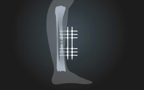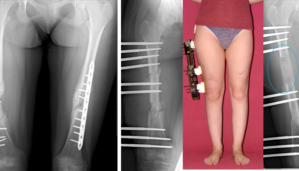
An infected nonunion is a fracture that fails to heal because infection has developed at the fracture site. A complex (recalcitrant) nonunion is one that has not healed despite one or more operations, where plates, nails or screws have broken or loosened, or where there is bone loss or serious soft tissue damage. Unlike a straightforward nonunion, these situations usually need staged and comprehensive revision surgery: removing infected and non-viable bone, stabilising the fracture securely, repairing the soft tissues where necessary and rebuilding the resulting bone defect with techniques such as bone transport or the Masquelet technique.
This page has been written for patients and families facing a long and demanding course of treatment. The general definition of nonunion, how it differs from delayed union and the first-line treatment options are explained on our nonunion surgery page. Here we focus on the more complex situations in which initial treatment has not been enough.
What is a complex or infected nonunion?
A fracture that has not healed within the expected time and shows no sign of healing on its own is called a nonunion, or pseudarthrosis (a "false joint"). Some nonunions heal after a single well-planned operation. A nonunion is considered complex, however, if any of the following apply:
- It has failed to heal after one or more operations
- Plates, nails or screws have broken, loosened or moved
- There is infection at the fracture site: discharge, a sinus (a draining track to the skin) or microbiologically proven infection
- There is bone loss, or there are fragments of dead (non-viable) bone
- There is associated shortening, angulation or rotation
- The skin and soft tissues are in poor condition, or the circulation is compromised
In these fractures the problem rarely has a single cause; biological, mechanical and infective factors are usually present together.
Complex and infected nonunions are most common in the shin bone (tibia), mainly because the soft tissue covering its front surface is thin and fractures here are more often open. The thigh bone (femur), the upper arm (humerus) and the forearm bones can also be affected. High-energy injuries, open fractures and severe soft tissue damage at the time of injury are the main factors that increase the risk.
Why does a nonunion keep failing to heal?
For a fracture to heal, several conditions must be met at the same time: adequate stability at the fracture site, a good blood supply, bone-forming cells, a scaffold on which new bone can grow and the right growth factors. This framework, known in orthopaedics as the "diamond concept", also takes the patient's general health into account. A nonunion may persist because of:
- Mechanical factors: too much movement at the fracture, or the opposite, no load reaching it to stimulate healing; or an implant that is not strong enough for the fracture.
- Biological factors: a blood supply damaged by severe injury or repeated surgery, and bone loss.
- Infection: bacteria form a protective layer called a biofilm on implants and dead bone, which greatly reduces the effect of antibiotics.
- Patient factors: smoking, poorly controlled diabetes, poor nutrition, vitamin D deficiency, vascular disease and some medicines that can affect bone healing.
The first step towards a successful revision is to identify accurately which of these factors are involved. A further operation that does not address the underlying cause risks meeting the same problem again. Our article on the treatment and revision of complex fractures also discusses the factors that influence the outcome.
How is infection recognised? Symptoms and diagnosis
Sometimes infection is obvious, with discharge or a sinus. At other times it follows a "silent" (low-grade) course, showing itself only as persistent pain, mild redness, slow wound healing or a nonunion that cannot otherwise be explained. Diagnosis combines several tools:
- History and examination: whether the original injury was an open fracture, previous operations, wound problems and any history of discharge.
- Blood tests: inflammatory markers such as CRP and ESR can be helpful, but normal results do not rule out infection.
- X-rays and CT: show the state of healing, bone loss, fragments of dead bone (sequestra) and the condition of any implants.
- MRI and nuclear medicine scans: may be used in selected patients to show how far the infection extends.
- Deep tissue samples: multiple samples are taken from different areas during surgery to confirm the diagnosis and choose the right antibiotic.
Because antibiotics can affect the results of these samples, the treating team decides when antibiotics should be started; it is important not to start or stop antibiotics on your own. The general diagnosis and treatment of bone infection are covered in detail on our page on bone and soft tissue infections.
The core principles of revision surgery
Treating complex and infected nonunions usually calls for a team approach, with orthopaedic surgeons working alongside infectious disease specialists and, where needed, plastic surgeons. The key steps are:
- Radical debridement: removing infected and non-viable bone and soft tissue until healthy, bleeding bone is reached, and removing failed implants.
- Dead space management: filling the cavity left after debridement with antibiotic-loaded bone cement or absorbable carriers, which deliver high local concentrations of antibiotic.
- Stable fixation: external fixators are often preferred while infection is active; internal fixation may be used once the infection is under control.
- Soft tissue cover: covering the bone with healthy, well-vascularised tissue, using a tissue transfer (flap) if necessary.
- Targeted antibiotic treatment: systemic antibiotics chosen according to culture results, usually given over several weeks.
- Bone reconstruction: rebuilding the bone defect once infection is controlled or, in suitable cases, during the same operation.
Complex nonunions are often accompanied by shortening, angulation or rotation. The revision plan should include correcting these deformities as part of the same treatment; otherwise, even if the bone unites, the limb may not function as well as expected. The assessment of fractures that have healed crooked is covered on our page on deformity and malunion surgery.
Treatment may be carried out in one stage or in several; the choice depends on the extent of infection, the amount of bone loss and the condition of the soft tissues. Our article on the use of external fixators in nonunion explains their role in more detail.
Techniques for reconstructing bone loss
Bone transport
Bone transport is based on the principle of distraction osteogenesis described by Ilizarov. A segment of healthy bone is separated by a controlled cut and moved into the gap at about 1 mm a day using an external fixator or, in some situations, internal devices, while new bone forms behind it. When the transported segment reaches the far end, this "docking site" often needs a small additional procedure and bone grafting to help it unite. Bone transport is effective for large defects, but treatment lasts many months. The general principles of the method are explained on our page on Ilizarov and limb reconstruction surgery.
The Masquelet (induced membrane) technique
This is a two-stage method. In the first stage, a spacer made of bone cement, usually loaded with antibiotic, is placed in the cleaned defect and the bone is stabilised. The body forms a thin, well-vascularised membrane around the cement. In the second stage, usually a few weeks later, the cement is removed, the membrane is preserved and the pocket is filled with bone graft taken from the patient's own skeleton. The membrane helps to nourish the graft and allows it to turn into bone rather than being reabsorbed.
Acute shortening with lengthening elsewhere
When bone loss is limited, the bone ends can be brought together and compressed during surgery. The resulting shortening can then be corrected over time by lengthening through a controlled cut in a healthy part of the same bone. This gives direct contact between the bone ends, but how much shortening the soft tissues and blood vessels will tolerate has to be taken into account.
Bone grafts and vascularised bone transfer
Small defects are filled with the patient's own bone graft, most often taken from the pelvis. In selected situations, a vascularised transfer, such as a fibula moved together with its blood vessels using microsurgery, may be considered. Our article on bone grafts used for nonunion describes the options in more detail.
| Technique | Usually preferred for | Advantages | Points to consider |
|---|---|---|---|
| Bone transport | Medium to large bone defects, associated shortening or deformity | The gap fills with the patient's own new bone; deformity and shortening can be corrected at the same time | Long time in a fixator, pin-site care, extra procedure at the docking site |
| Masquelet technique | Medium-sized defects after infection has been cleared | May shorten time in a fixator; can be used with internal fixation | Two operations; a sufficient amount of graft is needed |
| Acute shortening and lengthening | Limited bone loss | Direct contact between bone ends | Limited by what the soft tissues and blood vessels will tolerate |
| Graft or vascularised bone transfer | Small defects or selected special situations | Can often be done in a single stage | Pain at the donor site; vascularised transfer requires microsurgery |
The treatment journey and living with a fixator
Treatment of a complex nonunion often takes many months and sometimes more than a year. Active involvement from the patient and family makes a real difference:
- Pin and wire site care: if an external fixator is used, cleaning the pin sites regularly as you have been shown reduces the risk of infection.
- Physiotherapy: keeping joints moving and muscles strong has a direct effect on function once treatment is over.
- Nutrition and smoking: adequate protein intake and stopping smoking support bone healing.
- Pain management and sleep: regular contact with the treating team makes pain easier to keep under control.
- Psychological support: a long course of treatment can be exhausting, and professional support when needed can help you stay on track.
- Regular follow-up: healing and infection are monitored with X-rays and blood tests.
Planning work or school arrangements, getting around at home and transport to appointments before treatment begins makes this long process more manageable.
Risks and realistic expectations
Treatment of complex and infected nonunions always involves a degree of uncertainty, so it is important to discuss expectations openly from the outset:
- Recurrence of infection: bone infection can flare up again even years later, so long-term follow-up is needed.
- Persistent nonunion: further operations may be needed, particularly at the docking site.
- Pin-site infection, joint stiffness and muscle tightness
- Leg length difference, angulation or rotation
- Pain at the bone graft donor site
- Nerve or blood vessel injury
- Refracture: particularly in the period after a fixator has been removed.
In very rare cases, if infection and nonunion cannot be controlled despite repeated operations, or if continuing long treatment seems to offer little for quality of life, amputation and a prosthetic limb may need to be discussed honestly as an option. This is a decision made together with the patient, only after every alternative has been considered.
When should you seek advice?
An assessment by an orthopaedic surgeon is advisable if you have:
- Pain on weight-bearing, or a sense of movement at the fracture, months after fracture surgery
- Discharge from the surgical wound, a sinus opening on the skin, redness or recurrent fever
- Broken or loosened plates, nails or screws on follow-up X-rays
- A fracture that has not healed despite more than one operation
- Shortening, angulation or rotation of the limb after a fracture
If discharge is accompanied by a high temperature or rapidly spreading redness, seek medical care without delay.
Prof. Dr. Halil İbrahim Balcı's approach
Prof. Dr. Halil İbrahim Balcı is a faculty member in the Department of Orthopaedics and Traumatology at Istanbul University's Istanbul Faculty of Medicine. He worked at the Paley Institute in the USA as a research fellow (2014) and observer (2019) in Ilizarov techniques and deformity surgery, and is a co-editor of the Springer book "Basic Techniques for Extremity Reconstruction: External Fixator Applications According to Ilizarov Principles" (2018). He is a member of Turkish ASAMI and the Limb Lengthening and Reconstruction Society (LLRS). For complex nonunions, the treatment plan is built on a detailed review of previous operations, an assessment for infection and the patient's own expectations. Consultations are available in Turkish, English and French; please bring your previous imaging and operation notes when you request an assessment through the appointment page.
Key points
- A complex nonunion is a fracture that has not healed despite more than one operation, or one with failed implants, infection or bone loss.
- Successful revision depends on accurately identifying the mechanical, biological, infective and patient-related causes.
- In infected nonunion, the core principles are radical debridement, local and systemic antibiotics, stable fixation and good soft tissue cover.
- Bone loss can be reconstructed with bone transport, the Masquelet technique, acute shortening and lengthening, or grafts.
- Treatment is long and demanding, and long-term follow-up and realistic expectations are essential.