Deformity surgery is the branch of orthopaedics that measures and plans the correction of bones that have departed from their normal shape and alignment, and corrects them with a controlled bone cut (osteotomy) or, in children, with methods that guide growth. A deformity may involve bending (angulation), twisting about the bone's own axis (rotation), sideways shift (translation) or shortening, and these problems often occur together. Deformities can be congenital, developmental, metabolic, the result of injury or infection, and affect both children and adults.
A deformity of the leg is not just a question of appearance. When alignment is disturbed, the load on the hip, knee and ankle is distributed unevenly, which can cause pain and gait problems and, over the long term, contribute to osteoarthritis. This page explains the general principles of how deformities are analysed, the methods of correction and what treatment involves. The right treatment for any individual can only be decided after examination and imaging.
What is a bone deformity? The main types
Deformities are classified according to the plane in which they occur and their nature. A single bone may have more than one problem, and both the femur and the tibia may be affected.
| Type of deformity | What it means | Examples |
|---|---|---|
| Angular (seen from the front) | The bone bends inwards or outwards | Varus (bow legs), valgus (knock knees) |
| Angular (seen from the side) | The bone bends forwards or backwards | Procurvatum, recurvatum (knee bending too far backwards) |
| Rotational | The bone is twisted inwards or outwards about its own axis | Tibial torsion, increased femoral anteversion (in-toeing) |
| Translational | The bone fragments are shifted sideways relative to each other | A fracture that has healed out of line |
| Length | The bone is short, or the two sides differ | Leg length discrepancy |
Many deformities lie not exactly in the front or side view but in an oblique plane between the two. Rotational problems, meanwhile, are easily missed on standard X-rays; we discuss this in our article on rotational deformity and hidden bone twists.
Causes and symptoms of deformities
Main causes
- Congenital causes: limb differences such as fibular hemimelia or congenital femoral deficiency.
- Developmental causes: conditions affecting the growth plate, such as Blount's disease.
- Metabolic disease: conditions that disturb bone mineralisation, such as rickets, particularly X-linked hypophosphataemic rickets.
- Skeletal dysplasias: genetic conditions affecting bone development, such as achondroplasia.
- Injury: a fracture that has healed bent, rotated or short (malunion), or growth plate injuries in children. Post-traumatic deformities are covered separately on our malunion surgery page.
- Infection and tumours: bone infections that damage the growth plate, and tumour-related conditions such as multiple hereditary exostoses or Ollier disease.
- Neuromuscular conditions: conditions that upset muscle balance, such as cerebral palsy, which can lead in particular to rotational problems and foot deformities.
Symptoms
- a visible bend in the leg, with the knees held far apart (bow legs) or close together (knock knees)
- pain in the knee, ankle or hip, often concentrated on one side of the joint
- in-toeing or out-toeing, frequent tripping or falls, recurrent ankle sprains
- a feeling of the kneecap slipping, or pain at the front of the knee
- a limp and back pain caused by a leg length difference
- shoes wearing down faster on one side
How is a deformity analysed? The mechanical axis and CORA
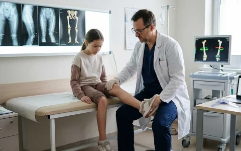
Accurate measurement is the first condition for a successful correction. Analysis begins with examination of leg lengths, joint movement, ligament laxity and the rotational profile, which is assessed with the patient lying face down. Imaging follows:
- Standing long-leg X-ray: a single radiograph showing both legs from hip to ankle, taken with the kneecaps facing forwards. This is the foundation of the analysis.
- Lateral X-rays: to assess forward and backward angulation.
- CT scanning: to measure rotation in the femur and tibia and, where needed, for three-dimensional planning.
On the long-leg X-ray, the mechanical axis is drawn first: a line from the centre of the hip joint to the centre of the ankle. Normally it passes through the centre of the knee or slightly to its inner side. If it passes well to the inner (medial) side of the knee there is varus (bow legs); if it passes to the outer (lateral) side, valgus (knock knees).
Next, the joint orientation angles are measured. For example, the angle between the lower joint surface of the femur and the mechanical axis (mLDFA) and the angle between the upper joint surface of the tibia and the mechanical axis (MPTA) are each normally about 87 degrees (85–90°). These angles show whether the deformity lies in the femur, the tibia, both, or at the level of the joint.
Loss of cartilage or ligament laxity inside the knee joint also affects leg alignment. The angle between the joint surfaces reflects this contribution and helps distinguish how much of the deformity arises from the bone and how much from the joint. A correction planned without making this distinction risks over-correction.
The key concept in analysis is the CORA (centre of rotation of angulation). This is the point where the axis lines of the upper and lower parts of the bone intersect, marking the apex of the deformity. When the correction is made at the level of the CORA, the bone is realigned both in angle and in axis. A correction made away from the CORA requires the fragments to be shifted as well; otherwise a new deformity may be created. Our article on the principles of deformity correction and CORA explains this in more detail, and computer-assisted planning is covered in 3D planning in deformity surgery.
Treatment options in deformity surgery
Non-surgical management and monitoring
Not every deformity needs an operation. Most bow legs and knock knees in young children are a normal stage of development and correct themselves with growth. In adults, a bone deformity will not be corrected by exercise, insoles or braces, although in mild cases physiotherapy, weight management and suitable insoles may ease symptoms. Surgery is considered when there is pain, progression or a risk of joint damage.
Guided growth (in children)
In children who are still growing, one side of a growth plate can be slowed temporarily with a small plate or screw, so that the bone straightens itself as it grows. Carried out through small incisions at the right time, this method may avoid the need for a larger osteotomy. Leg deformities in children are explained in detail on our page on childhood lower limb deformities.
Osteotomy: a controlled bone cut
- Opening wedge osteotomy: the bone is cut and opened on one side; the gap fills with bone on its own or is supported with a graft. It lengthens the bone slightly.
- Closing wedge osteotomy: a wedge of bone is removed and the ends are brought together. It shortens the bone slightly.
- Dome osteotomy: a curved cut allows large angular corrections without much change in length.
- Derotation osteotomy: for rotational deformities, the bone is cut and turned to the correct angle.
After the osteotomy the bone is held with a plate, an intramedullary nail or an external fixator. Specific techniques for correcting bow legs and knock knees around the knee in adults, such as high tibial osteotomy (HTO) and distal femoral osteotomy (DFO), are covered on our page on adult leg deformity correction.
Acute or gradual? Comparing methods of correction
A deformity can be corrected all at once during surgery (acute correction) or slowly over days and weeks after surgery using an external fixator (gradual correction). Both approaches have their place:
| Feature | Acute correction | Gradual correction |
|---|---|---|
| How it is done | Corrected in theatre and held with a plate or nail | A circular or hexapod frame is fitted, and correction is made with daily adjustments after surgery |
| Suitable for | Small to moderate deformities in few planes; healthy soft tissues | Large, multiplanar deformities; associated shortening; poor soft tissues or infection; situations with a high risk of nerve stretch |
| Fine-tuning | Cannot be changed after surgery | Can be fine-tuned according to follow-up X-rays |
| Lengthening | Limited | Can be combined with lengthening |
| Daily life | No external frame; shorter process | Frame and pin-site care; longer process |
The two approaches can be combined. In fixator-assisted nailing, for instance, the deformity is corrected in theatre with a temporary external fixator and then held with an intramedullary nail; in other patients, internal fixation is added once gradual correction is complete so that the frame can be removed early. The principles behind gradual correction are explained on our page on Ilizarov and limb reconstruction surgery.
The treatment process and recovery
- Planning: based on the examination and imaging, the angle to be corrected, the level of the osteotomy and the method of fixation are decided; digital or three-dimensional planning is used where helpful.
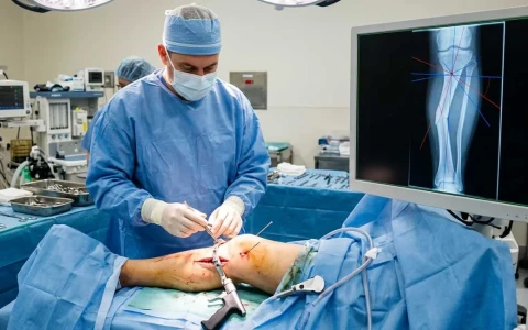
- Surgery: the osteotomy is performed under general or spinal anaesthesia and the bone is fixed with the chosen method. The hospital stay is usually a few days.
- Early phase: weight-bearing depends on the fixation used. Some plate-based corrections need a few weeks of partial weight-bearing, whereas circular frames usually allow early weight-bearing.
- If gradual correction is used: after a short latency period, the frame is adjusted every day according to the schedule provided, with regular X-ray check-ups.
- Bone healing: in adults the osteotomy usually takes a few months to unite; larger corrections and added lengthening take longer.
- Rehabilitation: physiotherapy restores joint movement, muscle strength and walking pattern. A return to sport is planned once the bone has fully healed.
How quickly the bone heals depends on age, the size of the correction and general health. Not smoking, eating a balanced diet, having vitamin D levels monitored by your doctor and following the weight-bearing instructions all support healing.
Plates and screws do not always have to be removed. They may be taken out in a separate operation once the bone has healed if they cause discomfort or, in children, if they might affect growth.
Possible risks and complications
Deformity surgery calls for careful planning and carries some risks:
- Under- or over-correction: the planned alignment is not fully achieved; with gradual correction this can usually be fine-tuned.
- Nerve problems: in particular, correcting a knock-knee deformity in the tibia all at once can stretch the nerve that runs around the outside of the knee. For this reason gradual correction, or releasing the nerve beforehand, is preferred in some cases.
- Compartment syndrome: a rise in pressure within the muscle compartments after surgery; rare but requiring urgent treatment.
- Delayed union or non-union of the osteotomy
- Infection: pin-site infection with external fixators; deep infection, which is less common, with internal fixation.
- Deep vein thrombosis, joint stiffness and irritation from implants
- Recurrence of the deformity: especially in growing children and in metabolic conditions such as rickets; in these patients the support provided by intramedullary nails can help reduce the risk of recurrence.
The likelihood of these risks varies from person to person with the size of the deformity, age, bone quality and general health, and should be discussed in detail before surgery.
When should you seek an assessment?
- You notice a bend in your leg that is getting worse or affects only one side
- You have persistent pain on one side of the knee or ankle
- Your leg has been left bent, rotated or short after a fracture
- Your child has bow legs that become more obvious or increase after the age of two, marked knock knees persisting beyond seven or eight, a deformity on one side only, or a limp
- In-toeing, frequent falls or a slipping kneecap are affecting daily life
Deformity surgery is usually a planned treatment, so there is time to talk through the options, expectations and risks in detail before deciding. After surgery, however, contact your treatment team without delay if you develop severe pain that keeps increasing despite pain relief, marked swelling and tightness of the leg, numbness in the foot or an inability to move your toes, or a fever.
Prof. Dr. Halil İbrahim Balcı's approach
Prof. Dr. Halil İbrahim Balcı studied deformity surgery and Ilizarov techniques at the Paley Institute in the USA as a research fellow in 2014 and as an observer in 2019, and has published more than 50 international papers on deformity, limb lengthening and paediatric orthopaedics. He is a faculty member in the Department of Orthopaedics and Traumatology at Istanbul Faculty of Medicine. For every deformity, the treatment plan is built on detailed measurement and planning, and whether correction should be acute or gradual is decided by considering each person's situation and expectations together. You can request a consultation through the appointment page.
Key points
- A deformity is angulation, rotation, translation or shortening of a bone, and these problems often occur together.
- Lower limb deformities disturb the load on the joints and can contribute to pain and, over time, osteoarthritis.
- Analysis is based on standing long-leg X-rays, the mechanical axis, joint angles and the CORA; rotation is measured with CT.
- Treatment ranges from monitoring and guided growth in children to osteotomy with acute or gradual correction.
- The right method is chosen individually after examination and imaging.