Paediatric orthopaedics is the branch of orthopaedics that diagnoses, monitors and treats conditions affecting children's bones, joints, muscles, ligaments and tendons, from birth until growth is complete at the end of adolescence. It covers a wide range of problems: developmental dysplasia of the hip and clubfoot in babies, in-toeing and bowed legs in toddlers, fractures in school-age children and spinal curvature in teenagers. Many of these findings are normal variations that correct themselves with growth; others can be treated with simpler methods when they are recognised early.
This page is intended as a roadmap for parents. It explains which problems are common at each age, how normal development is told apart from a condition that needs treatment, how children are assessed, the main treatment approaches and the warning signs that mean you should seek advice without delay. Specific conditions are covered in more detail on the related pages linked below.
Why is paediatric orthopaedics a separate field?
Children are not small adults. The growing skeleton has particular features that shape both diagnosis and treatment:
- Growth plates (physes): these areas of cartilage near the ends of long bones are where lengthwise growth happens. Damage to a growth plate can later lead to an angular deformity or a difference in leg length.
- Remodelling capacity: children's bones can straighten out some residual angulation after a fracture as they grow, which is why not every fracture in a child needs an operation.
- Timing: some treatments, such as small operations that guide growth, are only possible while the child is still growing, so assessment at the right time widens the options.
- Monitoring development: bone age, remaining growth and motor development are all taken into account when treatment decisions are made.
Children also cannot always describe what is wrong. A limp, not using one arm, holding back from play or general irritability may be the only sign of a musculoskeletal problem.
Common orthopaedic problems by age
| Age group | Common conditions | What parents may notice |
|---|---|---|
| Newborns and infants (0–1 year) | Developmental dysplasia of the hip, clubfoot (congenital talipes equinovarus), metatarsus adductus (a curved forefoot), torticollis | Uneven skin creases around the hips, limited spreading of the legs, a foot turned inwards and downwards, the head constantly tilted to one side |
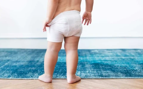
| Toddlers (1–4 years) | Physiological bow legs and knock knees, in-toeing, flexible flat feet, limping | A gap between the knees or knees that touch, frequent tripping, toes pointing inwards |
| School age (4–10 years) | Fractures, Perthes disease, growing pains, femoral anteversion (inward twist of the thigh bone) | A limp, hip or knee pain, a preference for sitting on the floor in a "W" position |
| Adolescents (10–18 years) | Scoliosis, slipped capital femoral epiphysis, Osgood–Schlatter disease, sports injuries, persistent bow legs or knock knees | Uneven shoulders or waistline, a limp with groin or knee pain, a painful lump below the kneecap |
Infancy
Developmental dysplasia of the hip (DDH) means that the hip socket and the head of the thigh bone have not developed in a well-matched way. It is more common in girls, first-born babies, babies born in the breech position and those with a family history of hip problems. Tight swaddling with the legs held straight can increase the risk, so hip-healthy swaddling that lets the hips bend and fall open is recommended. When DDH is picked up on ultrasound in the first months of life, it can usually be treated with simple measures such as a Pavlik harness. Our article on hip dysplasia and dislocation in children explains this in more detail.
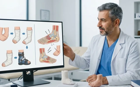
Clubfoot (congenital talipes equinovarus) is present from birth and causes the foot to point inwards and downwards. The first-line treatment today is the Ponseti method: the foot is corrected gradually with a series of weekly casts, most babies need a minor release of the Achilles tendon to complete the correction, and a foot abduction brace is then worn for several years to prevent relapse. We cover this in our clubfoot treatment guide.
School age and adolescence
Perthes disease usually appears between the ages of four and eight, when the blood supply to the head of the thigh bone is temporarily disrupted, causing a limp and pain in the hip, thigh or knee. Slipped capital femoral epiphysis (SCFE) typically affects adolescents: the head of the thigh bone slips at the level of the growth plate. Because it sometimes causes only knee pain it can be missed, and it needs urgent assessment. Adolescent idiopathic scoliosis, which is common in teenagers, may first be noticed as uneven shoulders, shoulder blades or waistline; when the child bends forward, one side of the back typically looks higher than the other.
Normal development or a problem that needs treatment?
One of the most common worries for parents is whether the shape of their child's legs and feet is normal. Many findings are simply a natural stage of growth:
- Bow legs: mild bowing is normal up to about the age of two.
- Knock knees: usually most noticeable around three to four years of age, and in most children the legs approach adult alignment by about seven or eight.
- In-toeing: in infants it usually comes from the forefoot, in toddlers from an inward twist of the shin bone (tibial torsion) and in school-age children from an inward twist of the thigh bone (femoral anteversion). Most cases improve with growth, and special shoes or insoles do not speed this up.
- Flexible flat feet: common in young children and usually need no treatment if they are not painful and an arch appears when the child stands on tiptoe.
- Growing pains: typically felt in both legs in the evening or at night, gone completely by the morning and never causing a limp.
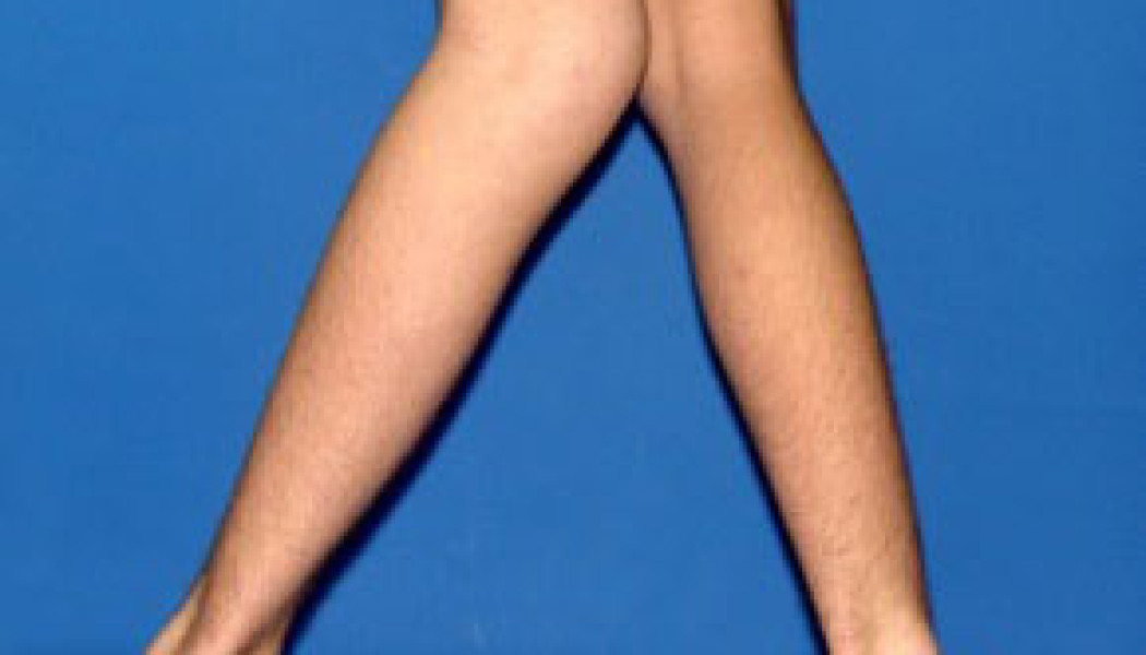
By contrast, a deformity that affects only one side, gets worse rather than better with age, causes pain or a limp, occurs with short stature or persists beyond the expected age range should be assessed in detail. Bowing and rotational problems of the legs in children are covered fully on our page on childhood lower limb deformities.
Congenital and rare conditions
Some children are born with, or develop during childhood, rarer conditions such as congenital limb deficiencies (for example fibular hemimelia or congenital femoral deficiency), congenital pseudarthrosis of the tibia (a shin bone that fails to unite), skeletal dysplasias such as achondroplasia, Ollier disease or multiple hereditary exostoses. Treatment for these children usually spans many years: it involves planned monitoring throughout growth, staged surgery where needed and teamwork with specialists in genetics, paediatric endocrinology and physiotherapy. These conditions are explained in detail on our page on rare childhood limb deformities.
In neuromuscular conditions such as cerebral palsy, spina bifida and muscular dystrophies, muscle imbalance can lead to hip displacement, foot deformities and spinal curvature. Orthopaedic follow-up for these children is planned together with paediatric neurology and physiotherapy.
Fractures and growth plate injuries in children
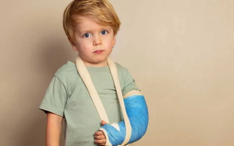
Because children's bones are more flexible, they break differently from adult bones. "Greenstick" fractures, in which one side of the bone breaks while the other bends, and "buckle" (torus) fractures, in which the bone simply buckles, are common. Most childhood fractures are treated in a cast or splint and heal faster than in adults. Fractures that involve a growth plate, however, can cause it to close early and lead to an angular deformity or a leg length difference, so they should be followed up for a while even after the bone has healed. Our article on common fractures in children is a useful guide. Leg length differences caused by growth plate damage or other conditions are covered on our page on leg length discrepancy and medical limb lengthening.
Examination and imaging tailored to children
Assessment begins with the pregnancy, birth and developmental history, the family history and watching how the child walks during play. The examination covers joint movement, leg lengths, rotational alignment, the spine and the nervous system. Avoiding unnecessary radiation is a guiding principle when choosing imaging:
- Ultrasound: the main method for checking the hips in the first months of life, with no radiation.
- X-rays: show bone structure and fractures, and standing full-length leg X-rays measure alignment.
- Bone age X-ray: usually of the left hand and wrist, used to estimate how much growth remains.
- MRI: uses no radiation and shows cartilage, growth plates and soft tissues; young children may need sedation to keep still.
- Low-dose imaging: where repeated images are needed, as in scoliosis follow-up, low-dose systems may be preferred when available.
Treatment approaches
Treatment in paediatric orthopaedics is often guided by a simple principle: the smallest effective intervention, carried out at the right time. The main options are:
- Observation and regular review: for physiological variations and mild conditions, the child's growth is monitored.
- Physiotherapy and exercise: for muscle imbalance, some walking difficulties and sports-related problems.
- Casts, splints and braces: for example serial casts in the Ponseti method, the Pavlik harness for hip dysplasia and a spinal brace for scoliosis.
- Guided growth: a small plate or screw placed on one side of a growth plate gradually corrects an angular deformity while the child grows.
- Surgical correction: osteotomies that cut and realign the bone, procedures that reshape the hip socket, and lengthening and reconstruction with external fixators or lengthening nails.
The right approach depends on the diagnosis, the child's age, the growth remaining and the severity of the deformity, and is decided after examination and imaging. Even two children with the same diagnosis may need different treatment plans.
A roadmap for families
- Keep notes: write down when the problem started, what time of day it is worse and whether there is a limp; short videos of your child walking can be very helpful.
- Bring previous records: earlier X-rays, ultrasound and MRI images and reports, growth charts and birth details all make assessment easier.
- Prepare your questions: note what you want to ask about the diagnosis, treatment options, expected duration and how often follow-up is needed.
- Stick with the treatment: with casts, braces and exercise programmes, much of the benefit depends on how consistently the family carries them out.
- Keep follow-up appointments: some conditions change as the child grows, and regular review means any intervention can be timed correctly.
- Involve your child: age-appropriate explanations help children feel safe and cooperate with treatment.
When should you seek advice?
Ask for an assessment by an orthopaedic specialist if your child has any of the following:
- A limp or refusal to bear weight; if there is also a fever, the child needs to be seen the same day because of the possibility of joint infection
- Bone pain that wakes the child at night, affects one side only or is getting worse
- Uneven skin creases around the hips, a difference in leg length, or a baby whose legs are difficult to spread apart
- Marked bow legs that affect one side, are getting worse or persist after the age of two; marked knock knees that persist after seven or eight
- A teenager who limps with groin, thigh or knee pain
- Uneven shoulders, shoulder blades or waistline
- Loss of motor skills the child had already gained, or persistent toe walking after the age of three
Even when none of these signs is present, routine checks with your child's paediatrician remain important for monitoring the hips, walking development and posture. An orthopaedic review around the time a child starts school, or during a growth spurt, can answer parents' questions and set up a follow-up plan if one is needed. If you are worried about something, asking early rather than waiting often removes unnecessary anxiety.
Prof. Dr. Halil İbrahim Balcı's approach
Prof. Dr. Halil İbrahim Balcı is a faculty member in the Department of Orthopaedics and Traumatology at Istanbul University's Istanbul Faculty of Medicine. At the Paley Institute in West Palm Beach, USA, he worked as a research fellow in 2014 and as an observer in 2019, in deformity surgery, Ilizarov techniques, congenital bone anomalies and paediatric orthopaedics. He has more than 50 international publications on deformity, limb lengthening and paediatric orthopaedics. For children, the treatment plan is built around the child's growth, the goals agreed with the family and the findings of examination and imaging. Consultations are available in Turkish, English and French, and you can request one through the appointment page.
Key points
- Paediatric orthopaedics covers musculoskeletal problems from birth to the end of adolescence, with the effects of growth at its centre.
- Bow legs, knock knees, in-toeing and flexible flat feet are, at certain ages, part of normal development.
- Early diagnosis of conditions such as hip dysplasia and clubfoot allows simpler treatment.
- A limp with fever, pain that wakes a child at night and a teenager limping with groin or knee pain should be assessed promptly.
- Treatment ranges from observation, casts and braces to guided growth and surgical correction, and is planned individually for each child.