Rare childhood limb deformities include congenital limb deficiencies such as fibular hemimelia, tibial hemimelia and congenital femoral deficiency, as well as congenital pseudarthrosis of the tibia, skeletal dysplasias such as achondroplasia, Ollier disease and multiple hereditary exostoses. Each is uncommon, but each can have a profound effect on a child's growth, walking and future life. What they share is that the problem changes as the child grows, and treatment is usually a planned journey over years rather than a single operation.
This page explains what these conditions are, how they are diagnosed and which treatment options are available today, with parents in mind. For common problems such as bow legs, knock knees, in-toeing or flat feet, see our page on childhood lower limb deformities; for a broader overview of children's bone and joint care, see paediatric orthopaedics.
What are rare childhood limb deformities?
Some of these conditions arise while the limbs are forming in the womb (congenital limb deficiencies); others are caused by genetic changes that affect the growth plates or the development of cartilage. The table below summarises the main ones:
| Condition | Area affected | Typical features | Main principle of treatment |
|---|---|---|---|
| Fibular hemimelia | Fibula, tibia, foot | Short leg, unstable ankle, missing toes | Foot and ankle reconstruction, staged lengthening, or a prosthetic option |
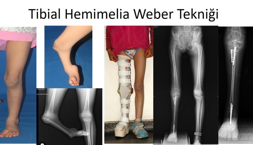
| Tibial hemimelia | Tibia, knee, foot | Severe shortening, unstable knee, inward-turned foot | Knee and foot reconstruction according to type, or a prosthetic option |
| Congenital femoral deficiency | Thigh bone, hip, knee | Short thigh, unstable hip and knee | Preparing the hip and knee, followed by staged lengthening |
| Congenital pseudarthrosis of the tibia | Shin bone | Forward and outward bowing, fracture that does not heal | Preventing fracture; surgery to achieve union |
| Skeletal dysplasias | Whole skeleton | Disproportionate short stature, bowed legs | Multidisciplinary care, correction of bowing and, in suitable patients, lengthening |
| Ollier disease and multiple exostoses | Around the growth plates | Shortening, angular deformity, bony lumps | Guided growth, correction, lengthening, long-term follow-up |
What is fibular hemimelia?
Fibular hemimelia means that the fibula, the slender bone beside the shin bone, is partly or completely absent from birth. It is the most common congenital deficiency of a long bone, yet it is still rare, and it usually affects one leg. The problem is rarely limited to the fibula itself; other features are often present:
- a short tibia that bows forwards
- an unstable ankle, with the foot drifting outwards (equinovalgus)
- fused bones in the foot and missing toes on the outer side
- a tendency to knock knee and deficient cruciate ligaments
- shortening of the thigh bone as well (associated femoral deficiency)
Classifications such as Achterman–Kalamchi and Paley guide the treatment plan; the Paley classification is based mainly on the condition of the ankle and the joint beneath it. In the past, amputation at ankle level followed by a prosthesis was common in severe cases. Today, in suitable children, the foot and ankle can be reconstructed, bowing corrected and the difference in leg length made up by staged lengthening during growth. Amputation with a prosthesis nevertheless remains a functional and valid choice for some children and families; the decision is made together, based on the foot, the predicted length difference and the family's preferences. More detail is available in our article on fibular hemimelia: diagnosis and Paley method treatment.
Tibial hemimelia
Tibial hemimelia is a partial or complete absence of the shin bone (tibia) and is much rarer than fibular hemimelia. Marked shortening, an unstable knee or limited knee bending, and an inward-turned foot (equinovarus) are typical. Because some families show inheritance and associated findings such as hand differences, genetic assessment is recommended.
Treatment depends on which part of the tibia is present (the Jones classification). If the upper tibia is present, the fibula can be joined to it to create a stable leg, the foot can be centred and, if necessary, the ankle fused, followed by lengthening. In the severe types where the tibia is completely absent, knee disarticulation with a prosthesis used to be the standard approach. In selected children, surgical techniques that aim to reconstruct a knee joint are now also used, but they involve a long, staged process and results vary from child to child. Both routes are discussed with the family in detail.
Congenital femoral deficiency (proximal femoral focal deficiency)
Congenital femoral deficiency means the thigh bone is short or incompletely formed from birth. At the mild end there is simply a short but solid femur; at the severe end, the part near the hip has not developed at all (proximal femoral focal deficiency). A reduced neck angle at the hip (coxa vara), a shallow socket, absent cruciate ligaments and an unstable kneecap are common, and many children also have fibular hemimelia in the same leg.
When a leg is lengthened, the hip and knee come under tension, and if these joints are unstable the risk of dislocation during lengthening rises. In suitable children, preparatory operations that stabilise and realign the hip and knee are therefore performed first, and lengthening is then planned in several stages during growth. For very severe deficiencies, an extension prosthesis or a rotationplasty, in which the ankle takes over the role of the knee, can also give good function. The right route depends on the type of deficiency, the state of the joints and the family's expectations.
Congenital pseudarthrosis of the tibia
Congenital pseudarthrosis of the tibia is a rare condition in which the shin bone bows forwards and outwards in infancy, breaks easily at that point and does not heal on its own once it has broken. Many affected children have neurofibromatosis type 1, a genetic condition associated with light brown skin patches. Not every bowed shin in a baby is this condition: bowing backwards and inwards (posteromedial bowing) usually straightens with time, although it may leave a difference in leg length.
Before a fracture occurs, a protective brace is used to shield the bone. Once a fracture has happened, surgery is needed: the abnormal tissue is removed, bone graft is applied and the bone is stabilised with an intramedullary rod and/or a circular external fixator. Some current techniques aim to strengthen healing by creating a broad bony bridge between the tibia and fibula. Even after union, the risk of refracture persists until growth has finished, so long-term protection and follow-up are needed.
Skeletal dysplasias, Ollier disease and multiple exostoses
Achondroplasia and other skeletal dysplasias
Skeletal dysplasias are genetic conditions that affect the development of bone and cartilage. The most familiar, achondroplasia, occurs in roughly 1 in 20,000 to 30,000 births and usually arises from a new genetic change rather than being inherited. Marked shortening of the segments closest to the body, bow legs and an increased curve in the lower back are typical. In infancy, monitoring of the base of the skull and the spine by neurosurgeons and paediatricians is important, and growth-directed medical treatments delivered by paediatric endocrinologists are now available.
Orthopaedic care may include correcting leg bowing and, in suitable patients, lengthening of the legs and arms. Lengthening is a personal decision with medical, psychological and ethical dimensions; the child's own views, the family's expectations and a multidisciplinary assessment are all taken into account. Details of arm lengthening are on our page on upper limb lengthening and deformity correction. In other dysplasias, such as hypochondroplasia, multiple epiphyseal dysplasia and pseudoachondroplasia, joint and alignment problems are followed on similar principles.
Ollier disease (multiple enchondromatosis)
In Ollier disease, multiple benign cartilage tumours (enchondromas) develop inside the bones, usually affecting one side of the body more than the other. These cartilage areas interfere with the growth plates and can cause leg length difference and angular deformity. Correction and lengthening can often be carried out with external fixators. Because there is a risk in adulthood that one of the cartilage tumours turns into a malignant tumour (chondrosarcoma), new pain or growth should be assessed, and long-term follow-up matters.
Multiple hereditary exostoses
Multiple hereditary exostoses is an inherited condition in which many bony outgrowths (osteochondromas) develop near the growth plates. They can keep growing until growth has finished, and may cause forearm shortening and bowing, knock knees and ankle valgus, leg length difference and pressure on muscles, tendons or nerves. Treatment includes removal of troublesome osteochondromas, guided growth in children who are still growing and, where needed, lengthening or corrective osteotomy. A lump that keeps growing or becomes painful after growth has finished should be assessed, because of a low but real risk of malignant change.
Diagnosis, growth prediction and treatment planning
Some limb deficiencies can be seen on the detailed ultrasound scan in the second trimester of pregnancy. When a diagnosis is made before birth, learning about the condition and the treatment options at that stage can ease anxiety and make the next steps easier to plan.
After birth, assessment includes:
- Examination: leg lengths, movement and stability of the hip, knee and ankle, the shape of the foot, skin findings and general development.
- X-rays: to establish which bones are present and their shape and length; once the child is walking, standing long-leg X-rays are taken.
- MRI: can show cartilage structures that have not yet turned to bone, such as the femoral head.
- Genetic assessment: guides families in skeletal dysplasias, neurofibromatosis and inherited conditions.
- Predicting the difference at skeletal maturity: in congenital length differences, the ratio between the short and long leg stays largely constant during growth, so calculations such as the multiplier method can estimate the final difference. This estimate is the key tool for deciding how many treatment stages will be needed.
The goals of treatment are a flat, comfortable foot, stable joints, equal or near-equal leg lengths at the end of growth and the ability to take part in activities appropriate for the child's age. Reaching them usually means combining reconstructive surgery in early childhood, lengthening repeated at intervals during growth and, where appropriate, slowing growth in the longer leg at the right time. The general principles of treating leg length discrepancy are explained on our medical limb lengthening page, and options specific to children in our article on leg length discrepancy in children.
The treatment journey, risks and the family's role
Treating a rare deformity is a long journey. A child may need several operations and may spend weeks or months in an external fixator. Physiotherapy, school arrangements, psychological support and the family's active involvement are as important as the surgery itself. Frame adjustments and pin-site care are usually taught to the family.
The main risks families should know about are:
- pin-site infections with external fixators
- stiffness, partial dislocation or dislocation of the hip or knee during lengthening; preparatory surgery aims to reduce this risk
- new bone hardening too early or too slowly, and fracture or bending after frame removal
- temporary or lasting nerve problems caused by stretching
- deformity returning with growth and the need for further surgery
- refracture after union in congenital pseudarthrosis
These risks are discussed in detail before treatment begins. Families who share in the treatment plan and its goals are better placed to manage the process.
When should you seek an assessment?
- a short or absent limb seen on a pregnancy scan, so that you can be informed before birth
- a newborn with one leg clearly shorter, an abnormally shaped foot or missing toes
- forward and outward bowing of the shin in a baby, especially with light brown skin patches
- several hard lumps on a child's arms or legs
- short stature that is marked and disproportionate compared with other children of the same age
- a leg length difference or deformity that increases with growth
- new pain, a fracture or a change in walking in a child who already has a diagnosis
For more on uncommon bone conditions in children, see our article on rare bone diseases in children.
Prof. Dr. Balcı's approach to rare childhood deformities
Prof. Dr. Halil İbrahim Balcı is a faculty member in the Department of Orthopaedics and Traumatology at Istanbul University, Istanbul Faculty of Medicine. At the Paley Institute in the United States he worked as a research fellow in 2014 and as an observer in 2019, focusing on congenital bone anomalies, deformity surgery and paediatric orthopaedics. His work on lengthening in achondroplasia, including four-segment lower limb lengthening in children and bilateral humeral lengthening, has received awards at national and international congresses. For rare deformities, the treatment plan is built individually after examination and imaging, bringing together an accurate diagnosis, growth prediction and the family's preferences. Consultations are available in Turkish, English and French through our appointment page.
Key points
- Fibular hemimelia, tibial hemimelia and congenital femoral deficiency are congenital limb deficiencies that affect the leg at different levels.
- Congenital pseudarthrosis of the tibia, skeletal dysplasias, Ollier disease and multiple exostoses change with growth and need long-term follow-up.
- Predicting the leg length difference at the end of growth is the key tool for planning treatment stages.
- Reconstruction with staged lengthening and prosthetic options are both valid routes where appropriate; the decision is made with the family.
- Treatment is a long process; risks are discussed in advance, and outcomes vary from child to child.