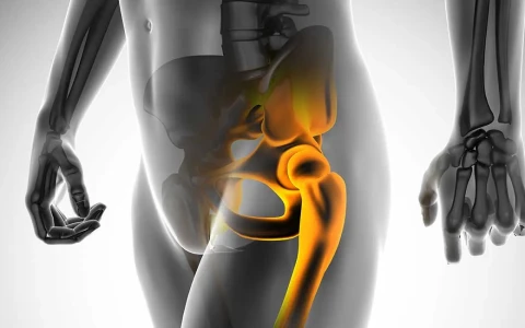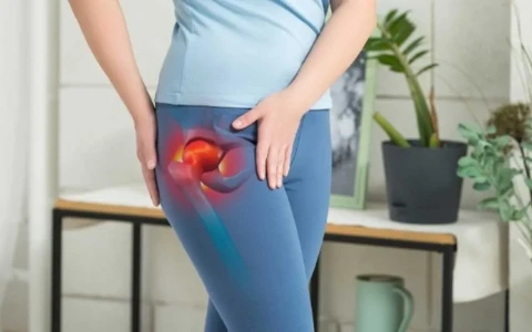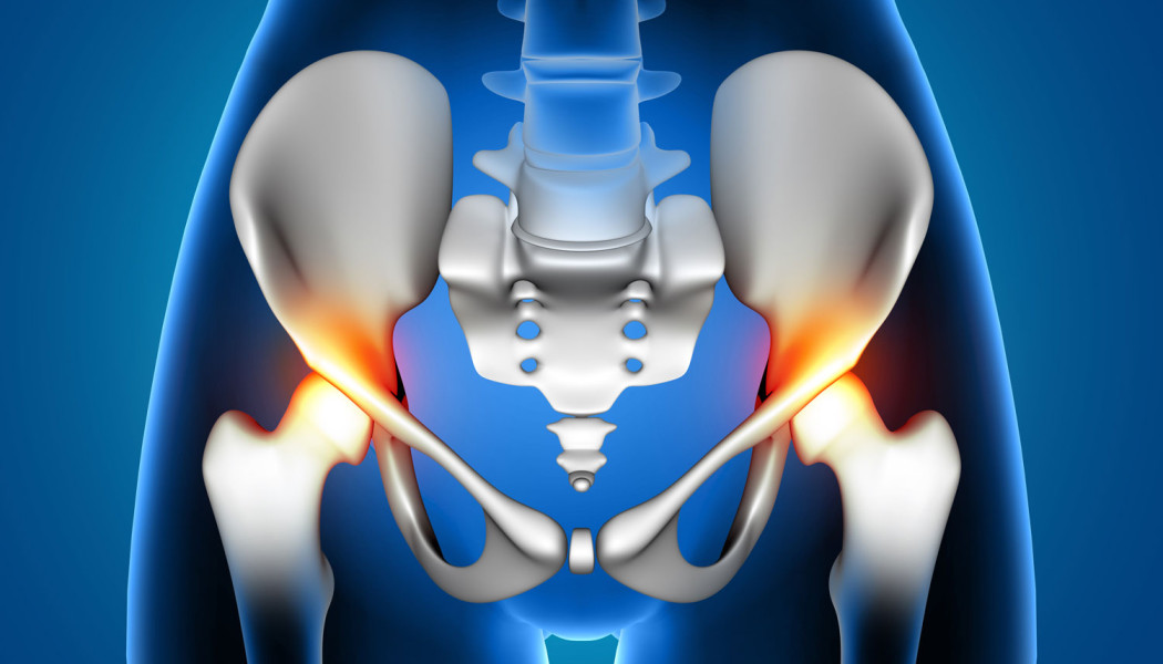
Hip osteoarthritis (coxarthrosis) is the gradual thinning and breakdown of the cartilage that covers the head of the thigh bone and its socket in the pelvis (the acetabulum). As cartilage is lost, the joint space narrows, bony spurs (osteophytes) form at the edges and the joint stiffens. The main symptoms are groin pain, loss of movement and difficulty walking. Hip osteoarthritis mostly affects people in middle and later life, but it can start much earlier in those with structural problems such as hip dysplasia or hip impingement.
Treating hip osteoarthritis is not just a matter of one operation. In the early stages, education, exercise and weight management can ease symptoms considerably; in younger people with a structural cause, joint-preserving surgery may be an option; and in advanced disease, hip replacement is an effective way to improve quality of life. This page explains the symptoms and stages, non-surgical treatment, joint-preserving options and how the decision for a replacement is made. For details of the operation itself, see our page on hip and knee replacement surgery.
What is hip osteoarthritis (coxarthrosis)?
In a healthy hip, smooth and resilient cartilage allows the ball of the thigh bone to turn freely in its socket and spreads load evenly. In osteoarthritis this cartilage wears away over the years. The bone beneath it, the joint lining (synovium), the joint capsule and the surrounding muscles are all involved as well. For this reason, osteoarthritis is now seen as a disease of the whole joint rather than simple "wear and tear".
The condition usually progresses slowly, and its course varies from person to person. Some people have marked changes on X-ray but only mild symptoms, while others have significant pain at an earlier stage. Treatment decisions are therefore guided more by symptoms and loss of function than by the appearance of the X-ray.
Causes and risk factors
When no specific underlying cause is found, the condition is called primary osteoarthritis; age, genetic predisposition and joint loading all play a part. When it develops as a result of another disease or a structural problem, it is called secondary osteoarthritis. Secondary causes are particularly important in younger patients:
- Developmental dysplasia of the hip: the socket does not cover the femoral head adequately, so load is concentrated on a small area and cartilage is damaged early. Mild dysplasia that went unnoticed in infancy may first show itself as arthritis in adulthood.
- Femoroacetabular impingement: differences in the shape of the femoral neck or the rim of the socket cause abnormal contact during movement, damaging the labrum (the rim of cartilage around the socket) and the joint cartilage.
- Avascular necrosis of the femoral head: loss of blood supply can make the femoral head collapse, and the joint may then deteriorate quickly.
- Childhood hip conditions: hips left misshapen after Perthes disease or slipped capital femoral epiphysis.
- Injury: irregularities left in the joint surface after fractures of the acetabulum or femoral head.
- Inflammatory arthritis and previous joint infection: rheumatoid arthritis, ankylosing spondylitis or a hip infection in childhood.
Excess body weight, physically heavy work and leg length difference can also make osteoarthritis more likely to develop or progress.
What are the symptoms of hip osteoarthritis?
- pain in the groin, sometimes at the side of the hip or the front of the thigh
- pain referred to the knee, so that some people think the problem lies in the knee rather than the hip
- "start-up" pain when standing after sitting for a while and during the first steps
- short-lived morning stiffness
- difficulty putting on socks or shoes, cutting toenails, or getting in and out of a car
- early loss of the ability to turn the hip inwards
- limping, shorter walking distances and difficulty on stairs
- in advanced stages, pain at night and at rest
Is the pain coming from the hip or the back?
Not all pain around the hip is due to osteoarthritis. A slipped disc or narrowing of the spinal canal in the lower back can send pain into the buttock and leg, and pain at the side of the hip is often caused by irritated tendons and bursae outside the joint (greater trochanteric pain syndrome). An inguinal hernia and other conditions can cause similar complaints. Pain from hip osteoarthritis is typically felt in the groin and is worse with hip movement, especially turning the hip inwards. The examination, with further tests where needed, tells these apart. Early warning signs are discussed in our article on early signs of hip osteoarthritis.
How is it diagnosed? Examination, X-rays and stages
Diagnosis is based on a careful history and examination, which looks at your walking pattern, hip range of motion, which movements bring on the pain, muscle strength and leg lengths. Imaging follows:
- Standing pelvic X-ray and a side view of the hip: show narrowing of the joint space, osteophytes, hardening of the bone under the cartilage and cysts. Shape problems that cause dysplasia or impingement can also be measured.
- MRI: shows early cartilage and labral damage, avascular necrosis and problems that are not visible on X-ray.
- CT scan: helps plan surgery when dysplasia or shape abnormalities are present.
- Blood tests: requested if inflammatory arthritis or infection is suspected.
X-ray findings are graded using systems such as Kellgren–Lawrence or Tönnis. The simplified table below summarises the stages; treatment, however, is always planned according to symptoms and not by stage alone.
| Stage | Typical X-ray appearance | Common symptoms | Main approach |
|---|---|---|---|
| Early | Mild narrowing of the joint space, small osteophytes | Intermittent groin pain, tiredness after long walks | Education, exercise, weight management; joint-preserving surgery in suitable younger patients |
| Moderate | Clear narrowing, osteophytes, hardening of the bone | Start-up pain, reduced movement, difficulty with daily tasks | Continue non-surgical treatment; discuss replacement if symptoms persist |
| Advanced | Joint space almost lost, cysts, change in the shape of the femoral head | Night pain, limping, marked loss of function | Hip replacement if symptoms impair quality of life |
Non-surgical treatment options
Non-surgical treatment is the foundation of care at every stage, and even when a replacement becomes necessary it helps you go into surgery in better condition. International guidelines regard exercise, education and weight management as core treatments.
- Exercise and physiotherapy: regular exercise that strengthens the hip muscles and maintains range of motion can reduce pain and improve function. Swimming, water-based exercise, a static bike and walking on level ground are usually well tolerated.
- Weight management: when you walk, the hip carries several times your body weight, so losing excess weight can reduce the load on the joint considerably.
- Adapting daily life: raised chairs and toilet seats, sock aids and using the handrail on stairs make everyday tasks easier.
- Walking stick: held in the hand opposite the painful hip, it reduces the load on the joint.
- Medication: painkillers and anti-inflammatory medicines are used under medical supervision, taking your general health into account.
- Joint injections: image-guided steroid injections can give short-term relief in some people. The evidence for hyaluronic acid and PRP in the hip is limited. Because joint injections given shortly before a hip replacement have been reported to increase the risk of infection, their timing should be planned with your surgeon.
Who is suitable for joint-preserving surgery?
Joint-preserving surgery aims to correct the structural problem that is driving the arthritis, so that you can keep your own hip for longer. It is most useful in younger and middle-aged people whose cartilage damage is still limited; once osteoarthritis is advanced, its benefit falls considerably.
- Periacetabular osteotomy (PAO): in dysplasia, the socket is cut free from the surrounding pelvis and repositioned so that it covers the femoral head better.
- Femoral osteotomies: used in selected patients whose femoral neck angle or rotation is disturbing the load on the joint.
- Hip arthroscopy or surgical hip dislocation: used in impingement to reshape the bone and repair a torn labrum. In hips with established arthritis, the benefit of arthroscopy is limited.
- Core decompression: may be used in suitable patients with early avascular necrosis, before the femoral head has collapsed.
How long these procedures can delay a replacement varies from person to person; this is discussed further in our article on whether joint preservation can delay hip replacement. For the symptoms and treatment of impingement, see our article on hip impingement syndrome.
When should a hip replacement be considered?
In a total hip replacement, the damaged joint surfaces are removed and replaced with an artificial socket and a new ball mounted on a stem inside the thigh bone. The decision is never based on the X-ray or on age alone. The following are usually considered together:
- pain that persists despite well-conducted non-surgical treatment
- loss of function that clearly affects sleep, walking, work and daily activities
- advanced joint damage on imaging that explains the symptoms
- general health that allows surgery, and realistic expectations
| Feature | Joint-preserving surgery | Hip replacement |
|---|---|---|
| Suitable patient | Usually younger, with a structural problem and largely preserved cartilage | Advanced osteoarthritis with insufficient benefit from non-surgical care |
| Aim | Keep your own joint and slow the progression of arthritis | Replace the damaged joint, reduce pain and improve function |
| Recovery | A few months on crutches when the bone needs to heal | Most people walk with an aid within the first days |
| Long-term consideration | A replacement may still be needed later | Revision may be needed over the years because of wear or loosening |
In suitable patients, modern hip replacements can function well for many years, but implant lifespan varies with age, activity level, bone quality and implant type. People who have a replacement at a younger age are more likely to need a revision operation later in life. Replacements made technically difficult by dysplasia, old fractures or leg length difference, as well as revision surgery, are covered on our page on complex and revision joint replacement.
Recovery after a replacement and possible risks
After a hip replacement, most people stand up with a physiotherapist on the day of surgery or the following day. The hospital stay is usually a few days. A frame or crutches are used for the first weeks, followed by a walking stick; the return to daily activities and driving varies from person to person. Depending on the surgical approach, you may be advised to take care with certain movements for a while.
Hip replacement is a common, planned operation, but like any surgery it carries risks:
- Infection: uncommon but serious, and it may require further surgery.
- Dislocation: the ball can come out of the socket, most often in the first months.
- Blood clots: measures are taken to prevent deep vein thrombosis and pulmonary embolism.
- Leg length difference and nerve problems: careful planning aims to keep these to a minimum.
- Fracture around the implant: during surgery or, in later years, after a fall.
- Loosening and wear: these may require revision over the years.
- Persistent pain or stiffness: a small number of people do not get the relief they hoped for.
When should you seek an assessment?
- groin or hip pain lasting more than a few weeks
- increasing difficulty putting on socks, tying shoelaces or getting into a car
- a limp or a clear reduction in how far you can walk
- pain that wakes you at night
- groin pain in a young adult with a history of hip dysplasia or dislocation in infancy
- being unable to bear weight after a fall, or sudden severe hip pain, which needs prompt assessment
- fever and feeling unwell together with hip pain, which needs urgent assessment for joint infection
Early diagnosis of hip dysplasia in childhood matters for the health of the hip in later life; see our article on hip dislocation in children.
Prof. Dr. Balcı's approach to hip osteoarthritis
Prof. Dr. Halil İbrahim Balcı is a faculty member in the Department of Orthopaedics and Traumatology at Istanbul University, Istanbul Faculty of Medicine. Because his academic work centres on deformity, limb lengthening and paediatric orthopaedics, structural problems that predispose to osteoarthritis, such as dysplasia, old fractures or leg length difference, are routinely included in the assessment. Together with you, he considers whether the pain truly comes from the hip, whether non-surgical treatment has been given a fair trial and what you want to be able to do in daily life; the treatment is chosen jointly after examination and imaging. Consultations are available in Turkish, English and French through our appointment page.
Key points
- Hip osteoarthritis is a breakdown of joint cartilage that causes groin pain, stiffness and difficulty walking.
- In younger patients, structural causes such as dysplasia and impingement should be looked for.
- Exercise, education and weight management are the basis of treatment at every stage.
- Joint-preserving surgery is an option for suitable patients whose cartilage is largely preserved.
- The decision for a replacement depends on persistent symptoms and loss of function, not the X-ray alone; outcomes vary from person to person.