Malunion means that a broken bone has healed, but not in the position it should have. The bone may have united at an angle, twisted around its own axis, shortened, or with a step in the joint surface. Malunion surgery corrects this by cutting the bone in a controlled way (an osteotomy), moving it into the right position and holding it while it heals again. It is considered when the malunion causes symptoms or is likely to overload the neighbouring joints over time.
Not every malunion needs an operation. Children's bones can remodel some angulation as they grow, and many adults live with small deviations without any loss of function. This page focuses on malunion after a fracture: which symptoms matter, how it is measured, when surgery is worth considering and what recovery involves. For congenital and developmental deformities and the principles of deformity analysis, see our page on deformity surgery.
What is a malunion and what types are there?
As a fracture heals, the bone ends are bridged by a callus that gradually hardens. If the fragments are not held in good alignment during this period, the bone still unites, but in a different shape. This is what separates malunion from nonunion: in a nonunion the bone ends fail to join; in a malunion they join in the wrong position. Occasionally both are present.
A malunion can involve one or more of the following components:
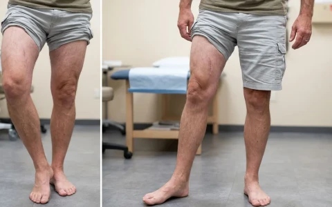
- Angulation: the bone has healed bent, either inwards (varus) or outwards (valgus) when seen from the front, or forwards or backwards when seen from the side.
- Rotation: the bone has healed twisted along its long axis. The typical sign is a foot that points noticeably further in or out than the other one, and it is easily missed on plain X-rays.
- Shortening: the limb is shorter because the fragments overlapped or because bone was lost.
- Translation: the fragments have healed shifted sideways relative to each other.
- Intra-articular step: when the fracture entered a joint, a step or gap left in the cartilage surface can lead to early wear of that joint.
Why does malunion happen?
There is rarely a single cause. Common contributing factors include:
- High-energy, multi-fragment fractures from road accidents or falls from a height, and injuries with bone loss
- Gradual loss of position in fractures treated in a cast or splint
- Incomplete alignment at the time of surgical fixation, or early loosening of an implant
- Putting full weight on the limb before the bone has healed sufficiently
- Conditions that slow bone healing, such as osteoporosis, smoking and diabetes
- In children, fractures that damage the growth plate (physis): if part of the growth plate closes early, the bone may grow increasingly angled and short
- Infection at the fracture site
Malunion can affect any bone, but is seen most often after fractures of the wrist (distal radius), forearm, femur, tibia and ankle. Two examples deserve particular attention: a rotational difference noticed after nailing of a femoral fracture, and an ankle fracture in which the fibula has healed short or rotated, which can disturb the fit of the ankle joint and lead to early arthritis.
What are the symptoms of malunion?
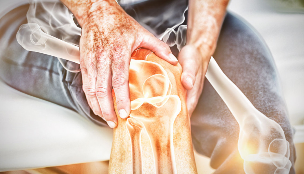
Symptoms depend on which bone is affected and in which direction it has healed. Some malunions stay silent for years; others affect daily life within months.
| Type of deformity | What you may notice | Why it matters in the long term |
|---|---|---|
| Angulation in the leg (bow-legged or knock-kneed) | Visible bowing, pain on one side of the knee or ankle, shoes wearing unevenly | Load concentrated on one half of the joint, with a risk of early arthritis |
| Rotational difference | A foot turned in or out, tripping, difficulty on stairs, pain around the kneecap | Uneven loading of the kneecap and hip |
| Shortening | Limping, low back pain, trouser legs hanging at different lengths | Long-term strain on the lower back and hip |
| Step in a joint surface | Joint pain, swelling, catching, reduced movement | Post-traumatic osteoarthritis |
| Forearm and wrist malunion | Difficulty turning the palm up or down, weaker grip, wrist pain | Lasting restriction of hand and arm function |
Which malunions need surgery?
The decision depends not only on how large the deviation is, but also on your symptoms, age, activity level and the condition of the neighbouring joints. In the leg, the key concept is the mechanical axis: a line drawn from the centre of the hip to the centre of the ankle, which normally passes close to the middle of the knee. If a malunion shifts this line towards the inner or outer side of the knee, that part of the joint is overloaded year after year.
Acceptable limits vary from bone to bone and between published sources. In the tibia, for example, angulation beyond roughly 5 degrees seen from the front or 10 degrees seen from the side, a rotational difference of more than 10–15 degrees, or shortening of more than 1–1.5 centimetres is usually examined more closely. These figures are only a guide: two people with the same angle may have very different symptoms. In fractures that involved a joint, even a step of a few millimetres in the joint surface can be significant.
Corrective surgery is usually considered when:
- the malunion causes pain, limping or loss of function
- the mechanical axis is clearly displaced and there are early signs of overload in the knee or ankle
- a rotational difference noticeably affects walking and everyday activities
- forearm rotation is reduced enough to limit hand function
- in a child, angulation or shortening caused by growth plate damage is increasing over time
A malunion that causes no pain, does not affect function and is borderline on measurement can often simply be monitored with regular check-ups.
Is malunion different in children?
Growing bones can reshape themselves, a process called remodelling. Angulation close to a growth plate and in the plane of joint movement can improve considerably, especially in young children. Rotational differences and angulation at right angles to that plane usually do not, a typical example being cubitus varus after an elbow fracture. When a growth plate has been damaged, the deformity may even increase, so the remaining growth potential is always taken into account. Our page on childhood lower limb deformities explains how leg alignment problems are managed during growth.
How is malunion diagnosed? Examination and imaging
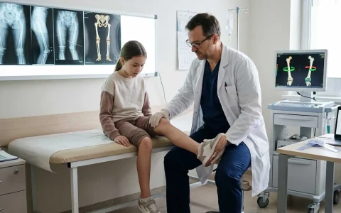
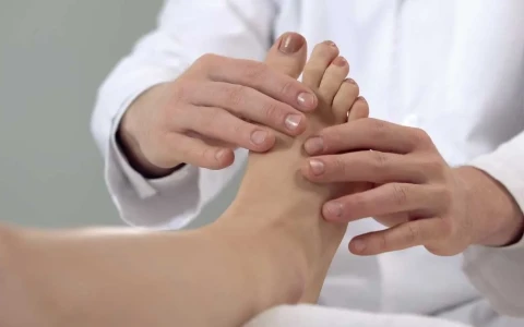
Assessment starts with your history: how the fracture happened, how it was treated, whether there was ever an infection and what troubles you now. The examination covers walking, leg lengths, joint movement, ligament stability and any rotational difference, which can be estimated by comparing hip rotation and the thigh–foot angle with you lying face down.
Imaging is tailored to the problem:
- Standing long-leg X-ray: a single image from hip to ankle, the basic tool for measuring the mechanical axis and leg length.
- Targeted X-rays: front and side views show the plane of the deformity and the state of healing. Views of the uninjured side are often taken for comparison.
- CT scan: measures rotation in degrees, shows steps in a joint surface and confirms whether the bone has fully united; 3D reconstructions help plan complex corrections.
- MRI: useful when the condition of cartilage, menisci or ligaments will influence the decision.
- Blood tests: markers of infection are checked if there has been an infection before, or if one is suspected.
Together, these findings define the plane, size and apex of the deformity. Rotational problems often need a dedicated assessment; our article on rotational deformity and hidden bone twists covers this in more detail.
Treatment options for malunion
Non-surgical measures
Non-surgical treatment does not change the shape of the bone, but it can ease symptoms and is sometimes all that is needed:
- a shoe raise or insert for leg length differences of up to 1–2 centimetres
- physiotherapy to improve muscle strength and joint movement
- weight management and adjusting activities that overload the joint
- pain relief as advised by your doctor and, where useful, a supportive brace
Corrective osteotomy
The cornerstone of surgical treatment is the corrective osteotomy: the bone is cut at a planned level, angle, rotation and length are restored, and the bone is fixed in its new position. A closing wedge osteotomy removes a wedge of bone; an opening wedge osteotomy opens the bone and fills the gap with graft; a dome osteotomy uses a curved cut; a derotation osteotomy corrects rotation alone. Carefully selected malunions inside a joint may be suitable for an intra-articular osteotomy, which is technically demanding. Old implants are removed during the same operation where necessary.
Acute or gradual correction?
The correction can be completed in one go during surgery (acute correction) and held with a plate or an intramedullary nail. Alternatively, it can be achieved gradually over days to weeks using an Ilizarov-type circular frame or a computer-assisted hexapod frame. When shortening also needs to be addressed, the gradual method allows the bone to be lengthened at the same time.
| Feature | Acute correction (plate or nail) | Gradual correction (external frame) |
|---|---|---|
| Typical situations | Moderate deformities in one or two planes, with healthy soft tissues | Large or multiplanar deformities, associated shortening, poor skin or soft tissue, previous infection |
| When the correction is achieved | During the operation | After surgery, through small daily adjustments |
| Nerves and blood vessels | Large sudden corrections can stretch them | Tissues are stretched slowly, which makes large corrections more controlled |
| Everyday life | No external device | Living with a frame for a period, with regular pin-site care |
| Fine-tuning afterwards | Limited | The correction can be adjusted according to follow-up X-rays |
The method is chosen after examination and imaging, based on the size of the deformity, bone quality, skin condition, previous operations and your circumstances. You can read more about circular frames on our Ilizarov and limb reconstruction page. If the malunion is combined with nonunion, bone loss or infection, the treatment plan becomes broader; these situations are covered on our page on complex and infected nonunion.
The surgical process and recovery, step by step
- Planning: measurements are made on the X-rays and, where helpful, the correction is simulated in advance on a computer or with 3D models.
- Preparation: a general health and anaesthetic assessment is carried out and conditions such as diabetes are optimised. Stopping smoking is strongly advised, because smoking can markedly slow bone healing.
- Surgery: the osteotomy is performed, the bone is moved into the planned position and fixed with a plate, nail or external frame. The length of the operation depends on the complexity of the deformity.
- Hospital stay: usually a few days, covering pain control, wound care and getting you moving again.
- If gradual correction is used: you and your family are taught how to make the frame adjustments, and the correction is checked at frequent visits.
- Healing phase: the osteotomy usually takes a few months to unite, and weight-bearing is increased step by step according to the fixation method and follow-up X-rays.
Recovery time varies with the bone, the size of the correction and the method used. After arm surgery people often return to daily tasks sooner; after leg surgery there is a period on crutches, and gradual correction with a frame takes longer overall. Physiotherapy is essential: early on it protects joint movement, later it rebuilds strength, balance and walking. Our article on recovery after malunion surgery explains what to look out for during this period.
Possible risks and complications
A corrective osteotomy is a planned operation, but like any surgery it carries risks:
- Infection: this may affect the wound or the bone. Pin-site infections are more common with external frames and can usually be controlled with dressings and treatment.
- Delayed union or nonunion of the osteotomy: more likely in smokers and in bones that have already had several operations.
- Nerve or blood vessel problems: mainly related to stretching during large, sudden corrections.
- Under- or over-correction, or loss of correction: a further adjustment or operation may be needed.
- Deep vein thrombosis: measures to reduce the risk of blood clots are taken after leg surgery.
- Joint stiffness and implant irritation: some people later need their plates and screws removed.
- Ongoing symptoms: if the joint cartilage is already damaged, pain may not resolve completely even when alignment is restored.
For these reasons, the decision to operate is a shared one, made after an open discussion of the expected benefits alongside the possible risks.
When should you seek an assessment?
After a fracture has healed, it is worth seeing an orthopaedic and trauma surgeon if you notice any of the following:
- visible bowing of the bone, or a clear difference in shape between the two sides
- a foot that points further in or out than it did before the injury
- a new limp, or the feeling that one leg is shorter
- increasing pain in the knee, ankle or wrist next to the healed fracture
- difficulty turning the palm of the hand up or down
- in a child, bowing after a fracture that increases over the following months
- redness, discharge or fever together with pain at the fracture site, which needs prompt assessment
Warning signs specific to the thigh and shin bones are described in our article on signs of femur and tibia malunion.
Prof. Dr. Balcı's approach to malunion surgery
Prof. Dr. Halil İbrahim Balcı is a faculty member in the Department of Orthopaedics and Traumatology at Istanbul University, Istanbul Faculty of Medicine. He worked at the Paley Institute in the United States as a research fellow in 2014 and as an observer in 2019, focusing on deformity surgery and Ilizarov techniques, and has contributed to more than 50 international publications on deformity, limb lengthening and paediatric orthopaedics. His assessment of a malunion is based on careful measurement, comparison with the other side and an honest conversation about your expectations; whether treatment is needed, and which method suits you, is decided after examination and imaging. Consultations are available in Turkish, English and French, and you can request one through our appointment page.
Key points
- Malunion means a fracture has healed angled, rotated, shortened or with a step in the joint surface; it is different from nonunion.
- Not every malunion needs surgery; the decision depends on symptoms, measurements and the condition of the nearby joints.
- Standing long-leg X-rays and, where needed, CT measurements are the basis for planning a correction.
- Correction can be done in one step with an osteotomy or gradually with an external frame; the choice is individual.
- Recovery time and outcomes vary from person to person; physiotherapy and regular follow-up are an important part of the process.