Femur (thigh bone) and tibia (shin bone) fractures can result in malunion if the anatomical alignment is not maintained during healing. This condition leads to functional and cosmetic problems that directly impact the patient’s quality of life. Especially when signs of femur malunion are observed, assessing the bone deformity carefully without causing additional joint damage is crucial.
Malunion is not simply about a crooked bone; it also involves shortening, rotational misalignment, and altered joint angles. Patients typically report limping, limb length discrepancy, or the kneecap pointing in an abnormal direction. Understanding these complex orthopaedic issues is an essential first step toward planning appropriate treatment.
What Is Malunion?
Malunion occurs when the fractured bone ends heal in an anatomically incorrect position. This can result from inadequate fixation, premature weight-bearing, or biological factors. Although the bone integrity is restored, angular or rotational deformities disrupt biomechanical balance. For a more comprehensive overview of malunion and treatment options, please visit what is malunion and how is it treated.
Malunions are generally classified into three main categories: angular deformities (bending), rotational deformities (twisting), and shortening. The impact of these deformities on bone structure is illustrated in the following comparative diagram showing normal versus pathological examples.
In orthopaedics, malunion is recognized not just as a cosmetic issue but as a functional problem that can lead to early osteoarthritis in adjacent joints (hip, knee, ankle). Therefore, detailed radiological examinations are necessary if symptoms persist after the fracture has healed.
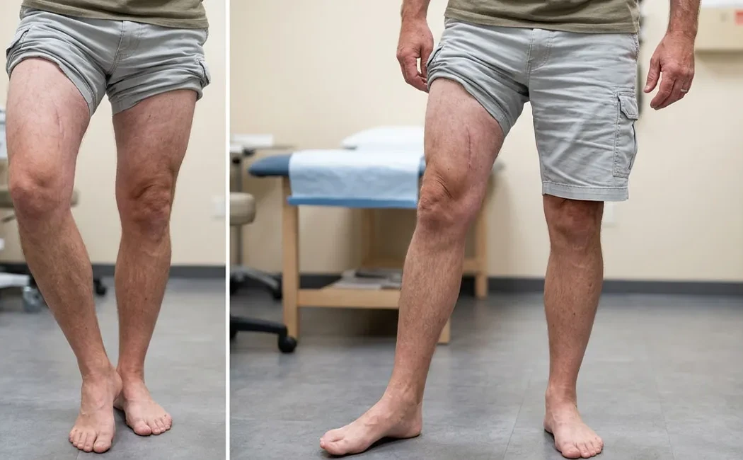
Signs of Femur Malunion
Since the femur is the longest and strongest bone in the body, malunions here produce noticeable symptoms. Signs of femur malunion typically manifest as alterations in the patient’s gait pattern. The most common findings include leg length discrepancy and visible deformity in the thigh region.
Patients often report:
- Limb Shortening: Caused by overlapping bone fragments during healing.
- Limping: Due to shortening or weakened hip abductor muscles.
- Knee and Hip Pain: Increased stress on joints from altered load transmission.
Complications during femur fracture healing sometimes overlap with nonunion issues. More details can be found in our article on nonunion in femur fractures.
Signs of Tibia Malunion
As a weight-bearing bone, even minor deviations in the tibia can cause significant problems in the knee and ankle joints. Tibia malunions often appear as an outward (valgus) or inward (varus) bowing of the lower leg. This condition can result in a “bow-legged” or “knock-kneed” appearance.
Symptoms include:
- Anterior Knee Pain: Related to disruption of the patellofemoral joint mechanics.
- Ankle Issues: Altered load distribution causes pain and limited mobility in the ankle.
- Cosmetic Deformity: Noticeable bowing or curvature in the lower leg.
Childhood fractures or growth plate injuries can also lead to tibial deformities later in life. Pediatric cases and deformity management are covered in detail on our lower limb deformities in childhood page. For corrective surgical options, see our lower limb deformity surgery resource.
Post-Fracture Limb Rotation (Rotational Deformity)
Rotational deformities are more difficult to detect than angular ones but can significantly impair patient function. Post-fracture limb rotation is identified by excessive inward or outward foot positioning during walking. This affects activities such as running, climbing stairs, or sitting cross-legged.
Detecting rotational deformities requires careful physical examination and CT imaging. Especially after femur fractures, errors in hip rotation angles influence the patella’s alignment. Similarly, tibial torsion disrupts ankle biomechanics. These complex cases may be associated with healing problems and can warrant evaluation for nonunion surgery.
What to Do If Signs of Malunion Are Noticed?
If you experience deformity, pain, or functional limitations after fracture treatment, consult an orthopaedic specialist promptly. Early-detected malunions can often be corrected with less invasive methods, whereas delayed diagnosis increases the risk of joint damage. Diagnosis usually involves physical examination, full-length leg X-rays, and CT imaging.
Treatment planning depends on the severity of the deformity and patient expectations. Surgical options include osteotomies (bone cuts for realignment) and external fixation systems like Ilizarov. For more information on surgery, visit malunion surgery. The postoperative period is equally important; details on recovery can be found in malunion surgery postoperative care.
If limb lengthening procedures are involved, our guide on post-limb lengthening surgery care may be helpful.
In conclusion, femur and tibia malunion, when treated timely and skillfully, can allow patients to regain mobility close to their original function. The key is not to ignore symptoms and to adhere to scientifically based treatment protocols.
This content is provided for general informational purposes only and does not constitute personal medical diagnosis or treatment advice. Always consult a qualified orthopedics and traumatology specialist for any health concerns. Do not delay seeking professional medical care based on this information.