A non-union is a fracture that has failed to heal in the expected time and in which the healing process has stopped. Because no solid bridge of bone forms between the fragments, pain, movement or a gradually increasing deformity may persist at the fracture site. Also known as pseudarthrosis ("false joint"), it can occur at any age but is more common in smokers, people with diabetes, after severe or open fractures, and in areas of bone with a relatively poor blood supply. Effective non-union treatment starts with understanding exactly why the bone has not healed, and then supplying what is missing.
Most fractures heal well with appropriate treatment; a non-union means that something has gone wrong along the way. Sometimes the bone has not been held stable enough, sometimes its biological capacity to heal is poor, and sometimes there is a hidden infection. This page explains the types of non-union, the causes, symptoms, diagnosis and treatment options. The right treatment for any individual is decided after examination and imaging.
What is a non-union, and how does it differ from delayed union?
The time a fracture takes to heal depends on the bone, the type of fracture and the person's health. Fractures of the arm, for example, tend to unite faster than those of the shin bone. So before concluding that a fracture "has not healed", the expected healing time for that particular injury is taken into account.
Normal fracture healing follows a sequence of stages. First, a blood clot (haematoma) and an inflammatory response form at the fracture; then a soft callus develops that links the bone ends together. This callus gradually hardens into bone, and finally the bone remodels over months or years towards its original structure. In a non-union, one link in this chain has broken.
| Feature | Delayed union | Non-union |
|---|---|---|
| Definition | Healing is slower than expected but still progressing | Healing has stopped; spontaneous union is unlikely |
| Timing | Usually 3–6 months after the fracture | Usually more than 6–9 months, with no progress in recent months |
| X-ray and CT | Bone bridging increasing, if slowly | No change on successive images; the fracture line remains open |
| Approach | Correcting risk factors, close follow-up, sometimes minor procedures | Surgery is usually needed |
A widely used definition describes a non-union as a fracture that is at least 6–9 months old and has shown no progress towards healing on successive check-ups, usually over the previous three months. However, the diagnosis may be made earlier if, for example, an implant has broken or there is a large gap between the bone ends.
Types of non-union
- Hypertrophic non-union: plenty of healing bone (callus) has formed around the fracture, but the ends have not joined. The biology is good; the real problem is insufficient stability, meaning the fracture is moving too much.
- Oligotrophic non-union: there is little callus, often because the bone ends are not in good contact or there is a gap between them.
- Atrophic non-union: there is almost no callus, and the bone ends have become thin and rounded. The problem is biological: the blood supply and healing cells are inadequate.
- Infected non-union: there is infection at the fracture site, and treatment has to address both the infection and the failure to heal.
- Synovial pseudarthrosis: a fluid-filled capsule resembling a joint has formed between the bone ends; this can develop in long-standing non-unions.
Why does non-union happen? Risk factors
Bone healing depends on four key elements: adequate stability, healing cells, a scaffold for new bone to grow on, and the growth factors that drive the process, all supported by a good blood supply and general health. Known in orthopaedics as the "diamond concept", this framework helps pinpoint why a fracture has not healed. The risk rises when any of these elements is missing:
- Patient factors: smoking and nicotine products, diabetes, vitamin D deficiency, poor nutrition, older age, marked obesity, vascular disease and long-term use of certain medicines that can affect bone healing.
- Injury factors: high-energy and comminuted (multi-fragment) fractures, open fractures, bone loss, severe damage to the surrounding soft tissues, and infection.
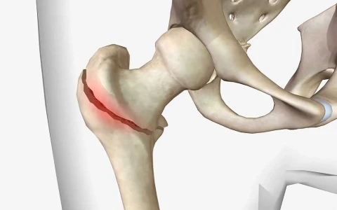
- Location: areas with a relatively poor blood supply, such as the scaphoid bone in the wrist, the neck of the femur, the lower part of the tibia and certain parts of the fifth metatarsal in the foot.
- Treatment factors: inadequate fixation, a gap left between the fracture ends, or early loosening or breakage of the implant.
Our article on non-union in femoral fractures and surgical approaches looks specifically at the thigh bone.
Symptoms of non-union
The signs of a non-union can be obvious or subtle. A fracture may not have healed if you notice:
- pain at the fracture site that persists for months, especially when bearing weight or using the arm
- abnormal movement, springiness or a clicking sensation at the fracture
- a gradually increasing bend or shortening of the arm or leg
- swelling and tenderness that do not settle
- a plate, screw or nail that breaks or loosens, often with a sudden onset of pain
- redness, warmth, fever or discharge from the wound, which may indicate infection
Some non-unions, particularly those held by a strong implant, cause very few symptoms for a long time and are picked up only on follow-up X-rays. That is why it is important to attend check-ups after a fracture.
How is non-union diagnosed?
The diagnosis is made by combining examination and imaging. To plan treatment properly, the aim is to answer not only "has it healed?" but also "why has it not healed?":
- Examination: tenderness and movement at the fracture, the state of the nearby joints, the skin and soft tissues, and the nerves and circulation.
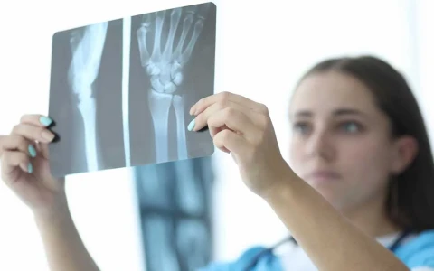
- X-rays: views from different angles, compared with earlier images; the condition of the implant is assessed at the same time.
- CT scanning: one of the most useful tests for showing whether bone has bridged the fracture, and often informative even when a plate or nail is present.
- MRI and nuclear medicine scans: used in selected cases to assess bone viability or infection.
- Blood tests: markers of infection (CRP, ESR, white cell count) and metabolic tests such as vitamin D, calcium, thyroid function and blood sugar.
- Long-leg X-rays: to measure any associated deformity or shortening in the lower limb.
If infection is suspected, several tissue samples are taken from the fracture site during surgery and sent for culture. The presence of infection changes the treatment plan considerably.
Non-union treatment: non-surgical and surgical options
Non-surgical approaches
For delayed union and some selected non-unions, the first step is to correct the factors that hold back bone healing: stopping smoking, controlling blood sugar, treating vitamin D deficiency under medical supervision and improving nutrition. The weight-bearing programme and any splint or brace may be adjusted. Bone stimulation methods such as low-intensity pulsed ultrasound or shock wave therapy may be tried in some patients, but the evidence for their effectiveness is limited, and they are no substitute for surgery if the fracture is mechanically unstable.
Surgical options
- Dynamisation: removing some of the locking screws from an intramedullary nail so that the fracture ends compress together as weight is put through the limb. This is used in suitable femoral and tibial fractures.
- Exchange nailing: removing the existing nail, reaming (widening) the bone canal and inserting a larger nail. The bone debris produced during reaming also helps to stimulate healing.
- Plate revision or augmentation: replacing failed fixation with a stronger compression plate, or adding a plate alongside an existing nail.
- Bone grafting and biological support: particularly in atrophic non-unions, the patient's own bone, usually taken from the wing of the pelvis, is placed at the fracture site. Where needed, bone harvested from the femoral canal, bone bank grafts, synthetic bone substitutes or bone marrow-derived products may be used.
- External fixation (Ilizarov): gradual compression of the fracture ends, with simultaneous correction of any deformity or shortening. This is particularly useful when the soft tissues are poor, infection is present or there have been several previous operations. The method is described on our page on Ilizarov and limb reconstruction surgery.
Correcting associated deformity and shortening
In long-standing non-unions the bone is often bent, rotated or shortened. If union is achieved without correcting these problems, the person may be left with a bone that has healed but is crooked and short. Planning therefore includes measuring alignment and length on long-leg X-rays, and correction can be carried out acutely during the non-union operation or gradually with an external fixator. If the shortening amounts to more than a few centimetres, bone lengthening can be planned as part of the same course of treatment.
Which bone graft is used in which situation is explained in our article on bone grafts used in non-union, and how stability and biology are tackled together is covered in biological and mechanical approaches in non-union surgery.
| Type of non-union | Main problem | Common approach |
|---|---|---|
| Hypertrophic | Insufficient stability; healing capacity good | Improving stability: compression plating, exchange nailing or compression with an external fixator; a graft is often unnecessary |
| Oligotrophic | Poor contact or a gap between the bone ends | Restoring contact and stable fixation; a graft if needed |
| Atrophic | Poor biology (blood supply and healing cells) | Removing non-viable tissue and providing stable fixation together with a bone graft or biological support |
| Infected | Infection, often combined with instability | Surgical clean-out (debridement), antibiotics guided by cultures and, usually, staged reconstruction |
Non-unions that have already been operated on several times, where implants have failed, or that are infected or involve bone loss need separate planning. This group, including techniques such as bone transport and the Masquelet technique, is covered on our page on revision surgery for complex and infected non-unions. If a bone has healed but in a bent, rotated or shortened position, the problem is malunion rather than non-union; this is discussed on our malunion surgery page.
The treatment process and recovery
- Preparation: risk factors are corrected as far as possible, and stopping smoking before surgery is strongly advised. Any suspicion of infection is investigated.
- Surgery: the non-union site is exposed, non-viable tissue is removed, any old implant is taken out if necessary, stable fixation is achieved with the chosen method and a graft is added if needed.
- Early phase: the hospital stay is usually a few days. Weight-bearing is set by the surgeon according to the type of fixation; some methods allow early weight-bearing.
- Follow-up: X-rays are taken at intervals, and CT is used if needed to judge whether the bone has united.
- Rehabilitation: physiotherapy maintains muscle strength and joint movement, and loading is increased gradually.
Patients can make a real contribution to the outcome. Avoiding smoking and nicotine products, eating a balanced diet with enough protein, following the weight-bearing instructions, doing the physiotherapy exercises regularly and attending every check-up are among the most important steps in supporting bone healing.
After revision surgery, union usually takes several months. The time depends on the type of non-union, the bone involved, general health and smoking. Some patients need more than one procedure to achieve union.
Possible risks and complications
Non-union surgery is often more complex than the original fracture treatment and carries some risks:
- persistent non-union and the need for further surgery
- infection at the operation site
- pain or tenderness where the graft was taken, for example at the pelvis
- injury to nerves or blood vessels
- stiffness of neighbouring joints and muscle weakness
- healing in a poor position, or a difference in leg length
- implant problems, deep vein thrombosis and the general risks of anaesthesia
The likelihood of these problems varies from person to person, and smoking, diabetes and the number of previous operations can be particularly important. The risks that apply to you should be discussed in detail before surgery.
When should you seek an assessment?
- Your pain has not eased, or is getting worse, three months or more after a fracture
- You have been told that follow-up X-rays show no progress in healing
- A plate, screw or nail has been found to be broken or loose
- Your arm or leg has developed a new or increasing bend or shortening
- There is redness, discharge or fever around the operation site (seek help promptly)
- Previous treatment for a non-union has not worked
Prof. Dr. Halil İbrahim Balcı's approach
Prof. Dr. Halil İbrahim Balcı is a faculty member in the Department of Orthopaedics and Traumatology at Istanbul Faculty of Medicine and obtained the European Board of Orthopaedics and Traumatology (EBOT) qualification in 2013. He co-edited the Springer book on external fixator applications according to Ilizarov principles (2018) and has served on the board of Turkish ASAMI since 2015. In non-union treatment, the first step is a detailed assessment of why the bone has not healed, looking at stability, biology, infection and patient factors, after which the treatment options are discussed with the patient. You can request a consultation through the appointment page.
Key points
- A non-union is a fracture in which healing has stopped; in delayed union, healing continues, if slowly.
- In hypertrophic non-union the problem is usually stability, in atrophic non-union it is biology; infection should always be looked for.
- Diagnosis combines examination, serial X-rays, CT and blood tests.
- Treatment may involve correcting risk factors, dynamisation, exchange nailing, plate revision, bone grafting or an external fixator.
- The right treatment depends on the cause of the non-union and is decided individually after examination and imaging.