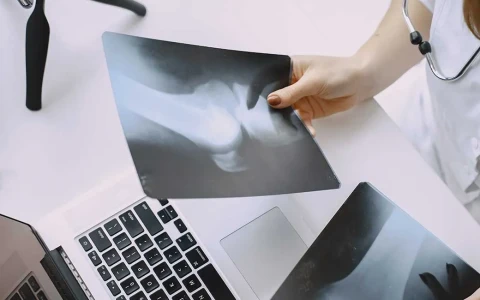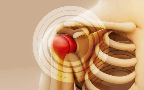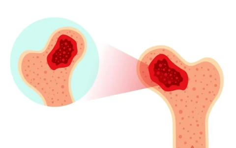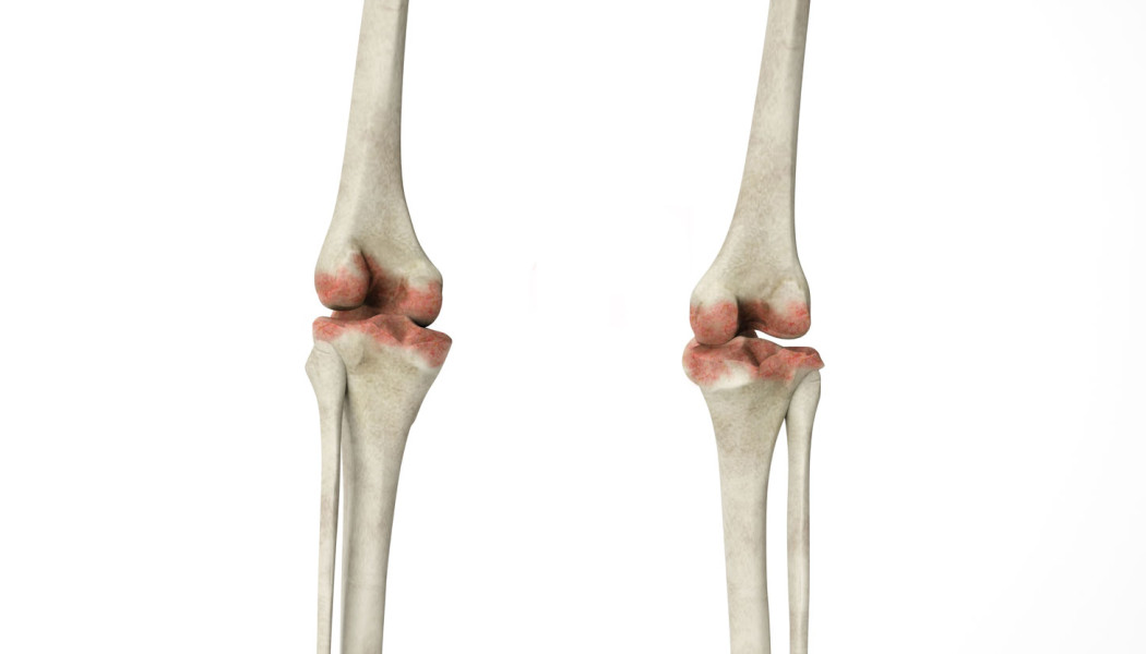
A bone tumour is an abnormal growth of tissue in bone caused by uncontrolled multiplication of bone or cartilage cells. Bone tumours can be benign or malignant. Most are benign: they do not spread to other parts of the body and usually do not threaten life. Malignant tumours that arise from the bone's own cells (bone sarcomas) are rare. In adults, a large proportion of malignant lesions found in bone are metastases, meaning cancers from organs such as the breast, prostate, lung, kidney or thyroid that have spread to the skeleton.
Bone tumours can occur at any age; some types are more common in children and young people, others in later life. An accurate diagnosis depends on combining examination, imaging and, often, a biopsy. Treating malignant tumours requires orthopaedic oncologists, medical oncologists, radiation oncologists, radiologists and pathologists to work together. This page explains the types of bone tumour, their symptoms, how they are diagnosed and the treatment options.
What is a bone tumour? Benign versus malignant
Whether a bone tumour is benign or malignant is the most important question in planning treatment. The table below summarises the general features, but a definite distinction can often be made only with imaging and examination of tissue.
| Feature | Benign bone tumours | Malignant bone tumours |
|---|---|---|
| Growth | Usually slow or static | Often rapid |
| Spread | Do not spread to other organs (with very rare exceptions) | Can spread, particularly to the lungs and other bones |
| X-ray appearance | Clear, smooth margins, often surrounded by a rim of hardened bone | Ill-defined margins, destruction of the outer bone, reaction of the periosteum and a possible soft tissue mass |
| Symptoms | Often none; frequently found by chance | Pain that increases over time and persists at rest and at night; swelling |
| Treatment | Monitoring, curettage, excision or needle-based procedures | Multidisciplinary treatment: surgery, chemotherapy and/or radiotherapy |
Primary tumours and metastases
Tumours that arise from bone tissue itself are called primary bone tumours. Lesions caused by cancer spreading to bone from another organ are bone metastases, and they are far more common than primary malignant bone tumours. For this reason, when a new destructive lesion is found in the bone of someone over about 40, the possibility of metastasis or multiple myeloma is always investigated.
Common types of bone tumour
Benign bone tumours and tumour-like lesions
- Osteochondroma: the most common benign bone tumour. It is a bony projection capped with cartilage that grows near a growth plate, and is usually noticed as a firm lump around the knee or shoulder. It generally stops growing when skeletal growth ends.
- Enchondroma: a cartilage tumour inside the bone, often in the bones of the hand and frequently found by chance.
- Non-ossifying fibroma: a very common lesion in children and teenagers that usually causes no symptoms and heals on its own over time.
- Simple and aneurysmal bone cysts: cavities filled with fluid or blood which, in children, can weaken the bone and lead to a fracture.
- Osteoid osteoma: a small tumour in young people that causes pain, especially at night; the pain is often markedly relieved by anti-inflammatory painkillers.
- Giant cell tumour: usually seen between the ages of 20 and 40 at the ends of bones, such as around the knee or the wrist. Although benign, it can damage surrounding bone and may recur after treatment.
- Fibrous dysplasia: a developmental condition in which normal bone is replaced by fibrous tissue, which can lead to bowing and fractures.
Malignant bone tumours
- Osteosarcoma: the most common primary malignant bone tumour in children and adolescents. It usually appears during the growth spurt, around the knee or in the shoulder.
- Ewing sarcoma: seen in children and young adults, commonly in the pelvis and the shafts of the long bones. Because it can cause fever and tiredness, it is sometimes mistaken for a bone infection.
- Chondrosarcoma: a tumour of cartilage origin, mostly affecting adults over 40 in the pelvis, the femur and around the shoulder.
- Multiple myeloma: a cancer of the plasma cells in the bone marrow, seen in older adults and treated by haematologists. The orthopaedic role is to prevent and treat fractures.
- Bone metastases: the most common cause of malignant bone lesions in adults, particularly in cancers of the breast, prostate, lung, kidney and thyroid.
The features of bone tumours in children and the importance of early diagnosis are covered in our article on bone tumours in children: symptoms and early diagnosis. For other bone and joint problems in children, see our paediatric orthopaedics page.
Symptoms of a bone tumour and when to see a doctor
Many benign bone tumours cause no symptoms at all and are discovered by chance on an X-ray taken for another reason. When symptoms do occur, the most common is pain. Bone tumour pain usually increases over time and persists at rest and at night, which distinguishes it from muscular aches that come on with exertion and settle with rest.
You should see an orthopaedic and trauma surgeon if you have:
- bone pain that does not settle within a few weeks, gets steadily worse or wakes you at night
- a growing, firm swelling or lump in an arm or leg
- a fracture after a minor strain or a small fall
- in a child, one-sided pain that is still present in the morning, a limp or swelling around the knee
- unexplained fever, weight loss or tiredness together with bone pain
- new bone pain in someone who has previously been treated for cancer
- an osteochondroma that continues to grow or becomes painful in adulthood
The common "growing pains" of childhood usually affect both legs, come on in the evening or at night, are completely gone by the morning, and are not accompanied by a limp, swelling or abnormal findings on examination. Pain that does not fit this pattern should be assessed. Most of these symptoms turn out to have causes other than a bone tumour, but examination and, where needed, imaging are important for an accurate diagnosis.
How are bone tumours diagnosed?
- X-rays: the first and most important test. The location of the lesion, its margins and its effect on the bone often give strong clues as to whether a tumour is benign or aggressive.
- MRI: shows how far the tumour extends within the bone and into the surrounding soft tissues, and its relationship to nerves and blood vessels; it is the basis of surgical planning.
- CT: shows bone structure in detail and, for malignant tumours, is also used to scan the lungs.
- Bone scan or PET-CT: used to check whether the tumour is present elsewhere in the body (staging).
- Blood tests: general health, calcium, certain enzymes and, where appropriate, tests to look for myeloma or other cancers.
Why the biopsy matters and how it is planned
If the imaging shows clearly that a tumour is benign, a biopsy may not be needed. For lesions that look aggressive or cannot be diagnosed with confidence, a tissue sample is taken for examination by a pathologist. This is often done with a large-bore needle under CT or ultrasound guidance, and sometimes through a small operation.
The biopsy is one of the most critical steps in treating a bone tumour. Its site and route must be planned so that the biopsy track can be removed together with the tumour in any later operation. For this reason, when a tumour might be malignant, it is recommended that the biopsy is planned after all imaging is complete and together with the team that will carry out the definitive treatment.
Treatment options for bone tumours
Treating benign tumours
- Monitoring: many lesions that cause no symptoms and do not weaken the bone are simply checked at intervals.
- Curettage and filling: the tumour is scraped out from inside the bone and the cavity is filled with bone graft or bone cement. For tumours that tend to recur, additional techniques may be used to treat the cavity walls.
- Surgical removal: used for lesions such as osteochondromas that cause pain or press on a tendon or nerve.
- Image-guided procedures: destroying an osteoid osteoma with heat energy (radiofrequency ablation), and needle-based treatments for some cysts.
- Fracture prevention: protective fixation may be used when a lesion has markedly weakened the bone.
Treating malignant tumours
For malignant bone tumours, the treatment plan is agreed at a multidisciplinary tumour board attended by the relevant specialists. Treatment for osteosarcoma and Ewing sarcoma usually includes chemotherapy before and after surgery, and radiotherapy may also be used for Ewing sarcoma. Surgery is the mainstay of treatment for chondrosarcoma. The surgical aim is to remove the tumour in one piece together with a surrounding layer of healthy tissue (wide resection).
Today, limb-sparing surgery is possible for most bone sarcomas of the arms and legs in suitable patients. Where the tumour surrounds major nerves and blood vessels, or where saving the limb would leave it without useful function, amputation followed by an artificial limb may offer a better quality of life. This decision is always made after detailed discussion with the patient and family.
Treating bone metastases
In metastatic bone disease the aims are to relieve pain, prevent or treat fractures and preserve mobility. Treatment may include therapy for the underlying cancer, radiotherapy, medicines that strengthen bone and protective surgical fixation of bones at high risk of fracture. This is managed together with the oncology team.
Reconstruction after tumour removal
Restoring the bone and joint that have been removed with the tumour is essential for the function of the arm or leg. The method is chosen according to the site of the tumour, the length of bone removed, whether the joint can be preserved, and the person's age and lifestyle:
| Method | Description | Points to consider |
|---|---|---|
| Tumour prosthesis (modular endoprosthesis) | A metal prosthesis replaces the section of bone and joint that has been removed | Allows early movement; loosening, infection and revision may occur over time. Extendable prostheses can be used in growing children |
| Bone bank graft (allograft) | Donor bone from a bone bank is joined to the remaining bone | A biological solution; delayed healing at the junction, fracture and infection can occur |
| Vascularised fibula graft | The patient's own fibula is transferred together with its blood vessels | Living bone that can thicken over time; requires microsurgery |
| Bone transport and lengthening | The bone gap is filled with new bone generated gradually from healthy bone | Aims for biological repair with the patient's own living bone; requires an external fixator for a long period |
| Rotationplasty | In selected young children, the ankle is rotated and repositioned to work as a knee | A specialised option that can allow an active life; its appearance needs to be discussed in detail with the family |
When the joint can be preserved, only the middle section of the bone is removed (intercalary resection) and the gap can be filled with graft or bone transport. The principles of bone transport and lengthening are explained on our page on Ilizarov and limb reconstruction surgery. Our article on surgical resection and reconstruction techniques in bone tumours covers these methods in more detail.
Treatment process, rehabilitation and follow-up
- Assessment and staging: imaging, biopsy and the necessary tests are completed; for malignant tumours, the treatment plan is agreed at the tumour board.
- Treatment before surgery: where needed, chemotherapy or radiotherapy is given before the operation.
- Surgery: the tumour is removed and reconstruction is carried out with the chosen method. The length of the hospital stay depends on the extent of surgery.
- Rehabilitation: physiotherapy starts early, with the aim of regaining joint movement, muscle strength and walking. The weight-bearing programme depends on the type of reconstruction.
- Follow-up: for malignant tumours, both the operation site and the lungs are imaged every few months in the first years and then less often, over many years. Benign tumours that tend to recur are also checked regularly.
We describe what is involved in follow-up in more detail in our article on the postoperative follow-up process in bone tumours.
Possible risks and complications
- Infection: the risk is higher with large tumour prostheses and in patients receiving chemotherapy or radiotherapy.
- Wound healing problems: more common in areas that have received radiotherapy.
- Local recurrence: the tumour can return at the same site in some benign as well as malignant tumours, which is why regular follow-up matters.
- Prosthesis problems: loosening, wear, breakage or dislocation; revision surgery may be needed over time.
- Graft problems: delayed healing at the junction or fracture of the graft.
- Nerve and blood vessel injury, joint stiffness and loss of muscle strength.
- Leg length difference in children: if the growth plate is removed with the tumour, lengthening may be needed in later years.
Side effects of chemotherapy and radiotherapy are monitored and managed by the oncology team. The likelihood of these risks depends on the type and site of the tumour, the treatments used and the person's general health, and should be discussed in detail before treatment.
Prof. Dr. Halil İbrahim Balcı's approach
The diagnosis and treatment of malignant bone tumours should always be planned through a multidisciplinary team decision. Prof. Dr. Halil İbrahim Balcı is a faculty member in the Department of Orthopaedics and Traumatology at Istanbul Faculty of Medicine, and his academic work focuses on limb reconstruction, bone lengthening and deformity surgery. His work at the Paley Institute in the USA and the Springer book on Ilizarov principles that he co-edited (2018) are closely related to the methods used to treat bone loss, leg length differences and deformities that can follow tumour surgery. Where needed, assessment is carried out in collaboration with the relevant specialties. You can arrange a consultation through the appointment page.
Bone infections can sometimes mimic tumours; for more on telling the two apart and on how infections are treated, see our page on bone and soft tissue infections.
Key points
- Most bone tumours are benign; malignant tumours arising from bone itself are rare.
- In adults, a large proportion of malignant bone lesions are cancers that have spread from other organs.
- Pain that keeps increasing and persists at night, a growing lump or a fracture after a minor strain should be assessed.
- Diagnosis combines X-rays, MRI, CT and, where needed, a carefully planned biopsy.
- Treatment of malignant tumours is multidisciplinary, and limb-sparing surgery with reconstruction is possible in suitable patients.