Arm lengthening surgery corrects shortening of the upper arm bone (humerus) or of one or both forearm bones (radius and ulna) by cutting the bone in a controlled way and then lengthening it gradually over several weeks. Upper limb deformity correction covers the straightening of bowing and rotational problems in the arm, elbow, forearm and wrist using corrective osteotomies. These operations are most often considered in skeletal dysplasias such as achondroplasia, in multiple hereditary exostoses, in Madelung deformity, and after childhood fractures or infections that damaged a growth plate.
Shortening is approached differently in the arm and the leg. In the leg, a difference of a few centimetres affects walking and the lower back; in the upper arm, a similar difference often causes no functional problem at all. In the forearm, however, the relative length and shape of the two bones directly determine how well the palm can turn up and down and how healthy the wrist remains. Treatment decisions in the upper limb are therefore driven by how the arm and hand work in daily life rather than by the measured shortening alone.
Why do shortening and deformity matter in the upper limb?
The main job of the upper limb is to place the hand where it is needed, reliably and comfortably. The shoulder, upper arm, elbow, forearm and wrist are links in that chain. A short humerus reduces the area the hand can reach: reaching behind the head, to the back, to the feet, or to the lower body for personal hygiene can become difficult. This is the most common reason people with achondroplasia ask about arm lengthening.
In the forearm, the radius and ulna are linked by two joints, one near the elbow and one at the wrist (the proximal and distal radioulnar joints). When we turn the palm up (supination) or down (pronation), the radius rotates around the ulna. If one bone stays shorter than the other or grows bowed, forearm rotation can be restricted, the wrist can tilt sideways and the head of the radius can dislocate at the elbow. Elbow deformities change the appearance of the arm and, in some people, cause instability or nerve symptoms.
When is arm lengthening or deformity correction needed?
Achondroplasia and other skeletal dysplasias
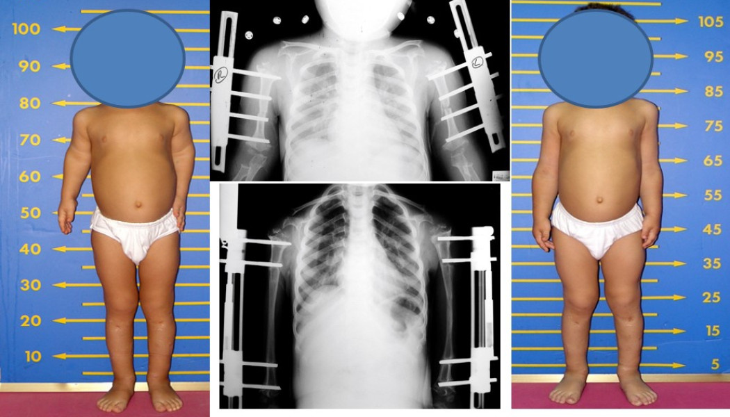
Achondroplasia is a genetic skeletal dysplasia that affects the growth plates; the segments closest to the body, the upper arms and thighs, are particularly short. A short upper arm can make dressing, reaching high shelves and toileting hygiene difficult. In suitable patients, bilateral humeral lengthening aims to extend the reach of the hands and increase independence in daily life. It is usually planned during childhood or adolescence, alongside the lower limb lengthening programme. Many people with achondroplasia cannot fully straighten the elbow; lengthening does not change this, and it is assessed separately. Some children now also receive growth-directed medical treatment from paediatric endocrinologists, and surgical plans take this into account.
Multiple hereditary exostoses (multiple osteochondromas)
In this inherited condition, bony outgrowths called osteochondromas develop near the growth plates. In the forearm, the growth plate at the wrist end of the ulna is affected most often: the ulna stays short, the radius bows, the wrist drifts towards the little-finger side and, in some children, the radial head dislocates at the elbow. Treatment can combine removal of troublesome osteochondromas, gradual lengthening of the ulna, corrective osteotomy of the radius and, in growing children, guided growth, in which one side of a growth plate is temporarily slowed down.
Madelung deformity
Madelung deformity develops when the inner, palm-side part of the growth plate at the wrist end of the radius does not grow properly. It typically appears in adolescent girls, is often bilateral, and in some cases is part of Leri-Weill dyschondrosteosis, which is linked to the SHOX gene. A prominent ulna at the back of the wrist, wrist pain and reduced movement are typical. In children who are still growing, release of an abnormal ligament (Vickers ligament) and freeing of the growth plate may be considered; once growth has finished, a dome-shaped corrective osteotomy of the radius, sometimes combined with a procedure on the ulna, is an option. Mild deformities that cause no pain may simply be monitored.
Shortening after growth plate damage, fractures or infection
Infection of the shoulder or arm in infancy, fractures through a growth plate, bone cysts next to a growth plate and conditions such as Ollier disease can all lead to marked shortening and bowing of the humerus or forearm. An injury to the arm nerves at birth (brachial plexus birth injury) can also leave the arm shorter and turned inwards. In all of these, the state of the growth plate and the remaining growth potential are assessed first.
Elbow deformities: cubitus varus and cubitus valgus
If a childhood fracture just above the elbow (a supracondylar fracture) heals in a poor position, the forearm angles inwards when the arm is held straight. This is called cubitus varus. It does not correct with growth and, although it is often mainly a cosmetic concern, it can cause elbow instability or nerve symptoms in some people. Cubitus valgus, which can follow a lateral condyle fracture that failed to heal, may gradually stretch the ulnar nerve over the years and cause numbness in the fingers. In both cases the treatment is a corrective osteotomy just above the elbow. Malunited fractures of the arm and forearm in adults are managed on similar principles.
Symptoms: what patients and parents notice
- one arm looking shorter than the other, or difficulty reaching because both arms are short
- difficulty dressing, brushing hair, reaching the back or feet, or with personal hygiene
- reduced ability to turn the palm up or down
- a wrist that tilts to one side, or a prominent bone at the back of the wrist
- a visible angle at the elbow, or an elbow that will not fully straighten or bend
- hard lumps on the arm or forearm (osteochondromas)
- numbness, tingling or weakness in the hand
- in children, a deformity that becomes more obvious with growth
Assessment: examination and imaging
The examination measures movement at the shoulder, elbow, forearm and wrist on both sides, and assesses grip strength, fine hand skills and nerve function. The everyday tasks that are difficult for you or your child are discussed one by one, because these define the goals of treatment.
- Comparative X-rays of both arms: showing the whole arm and forearm together with the shoulder, elbow and wrist. Bone lengths, the plane of the deformity, the length relationship between radius and ulna at the wrist and the position of the radial head are measured.
- CT scan: used for three-dimensional assessment of rotation and complex forearm deformities and, where appropriate, for patient-specific surgical planning.
- MRI: shows areas of premature growth plate closure (a physeal bar), cartilage and soft tissues.
- Hand and wrist X-ray for bone age: helps estimate how much growth remains, which guides the timing of surgery in children.
- Genetic assessment: can be helpful for families when a skeletal dysplasia, multiple exostoses or Madelung deformity is suspected.
Treatment options
Monitoring and supportive care
Mild shortening or bowing that does not affect function can usually be monitored. Physiotherapy and hand therapy help preserve joint movement, and splints or aids can make daily tasks easier, but none of these change the length or shape of the bone.
Bone lengthening (distraction osteogenesis)
Bone lengthening relies on the body's ability to fill a slowly widening gap between two bone ends with new bone. The bone is divided through a small incision and, after a waiting period of a few days, lengthening begins in several small steps a day using an external fixator or, in suitable patients, a lengthening nail placed inside the bone. In the upper arm the daily rate is usually around 1 millimetre; the forearm and smaller bones often need a slower pace. Once lengthening is complete, extra time is needed for the new bone to harden (consolidation). The humerus often tolerates proportionally more lengthening than the leg bones, and because the arm does not carry body weight, the hand can still be used during treatment. The biology of lengthening and the general principles of limb lengthening are explained on our medical limb lengthening page.
Corrective osteotomy
Bowing and rotation are corrected by cutting the bone at a planned level and moving it into the right position. The correction can be completed during surgery and held with a plate, wires or screws, or achieved gradually over days with an external fixator based on Ilizarov principles. In the forearm, lengthening and angular correction are often combined in a single treatment plan.
Guided growth and removal of osteochondromas
In children who are still growing, a small plate or screw placed on one side of a growth plate can temporarily slow growth on that side, so the bone gradually grows straighter. In multiple exostoses, troublesome osteochondromas can be removed as part of the same plan.
| Method | Typical uses | Key features |
|---|---|---|
| Lengthening with an external fixator | Humeral lengthening in achondroplasia, short ulna, shortening after growth plate damage | Lengthening and angular correction can be combined; pin-site care is needed |
| Internal lengthening nail | Selected adults with suitable bone dimensions | No external device; not suitable for every bone or every age |
| Acute corrective osteotomy | Cubitus varus, forearm malunion after fracture, Madelung deformity after growth has finished | Correction completed in theatre; large corrections need care with nerves |
| Guided growth | Wrist deformities in growing children | Small incisions; works over months and needs regular follow-up |
The treatment process, step by step
- Planning: the target length or correction is set with reference to the other side and to the person's daily needs. With families, timing around school and social life is planned as well.
- Surgery: the bone is cut and an external fixator or internal implant is fitted. With corrective osteotomies, fixation is completed in the same operation.
- Waiting period: if lengthening is planned, there is usually a pause of a few days to allow early healing at the bone cut.
- Lengthening phase: the patient or family makes the daily adjustments as they have been shown. X-rays are taken at regular visits, and nerve function and joint movement are monitored.
- Consolidation phase: after lengthening stops, the new bone is allowed to mature. This phase is often longer than the lengthening itself.
- Frame removal and rehabilitation: once the bone is strong enough, the fixator is removed and movement and strength are rebuilt with physiotherapy.
Recovery and rehabilitation
Keeping the shoulder, elbow, wrist and fingers moving during lengthening is as important as the bone work itself. As the bone lengthens, muscles, nerves and connective tissues are stretched too, so daily exercises and physiotherapy are an integral part of treatment. Children can usually keep going to school during lengthening; loose-fitting clothes over the frame and daily pin-site care soon become routine. The overall length of treatment depends on the amount of lengthening, the bone involved and the individual's healing, and varies from person to person. Our article on what to pay attention to after limb lengthening covers this period in more detail.
Possible risks and complications
Even with careful planning, arm lengthening and deformity correction carry risks. The main ones are:
- Pin-site infection: common with external fixators and usually controlled with dressings and treatment.
- Nerve problems: the radial nerve during humeral lengthening, and other nerves during forearm and elbow surgery, can be affected by stretching. Symptoms often settle when lengthening is slowed or paused, but further surgery is sometimes needed.
- Joint stiffness: movement at the shoulder, elbow or wrist may become restricted; regular physiotherapy reduces this risk.
- Early or delayed consolidation: the new bone may harden sooner than planned, or mature more slowly.
- Fracture or bending after frame removal: the new bone needs protecting until it is strong enough.
- Joint problems in the forearm: the radial head or the wrist joint can come under strain during ulnar lengthening.
- Recurrence: in growth-related conditions such as multiple exostoses and Madelung deformity, the deformity can return while growth continues, and further surgery may be needed.
Our article on complications in limb lengthening surgery explains how these problems are recognised and managed.
When should you seek an assessment?
An assessment by an orthopaedic and trauma surgeon is advisable if:
- short arms are limiting independence with dressing, reaching or personal hygiene
- your child's forearm or wrist is becoming more bowed with growth
- turning the palm up or down has become newly or increasingly restricted
- a child with multiple exostoses has a short forearm, a tilted wrist or a bony lump at the elbow
- an elbow has become visibly angled after a fracture, or feels unstable
- there is numbness, tingling or weakness in the hand
- an osteochondroma keeps growing or becomes painful after growth has finished, which should be assessed without delay
For congenital limb differences and rare skeletal conditions, see our page on rare childhood limb deformities.
Prof. Dr. Balcı's approach to upper limb lengthening and correction
Prof. Dr. Halil İbrahim Balcı is a faculty member in the Department of Orthopaedics and Traumatology at Istanbul University, Istanbul Faculty of Medicine. His work on bilateral humeral lengthening in achondroplasia received the first prize for external fixation oral presentations at the 24th National Turkish Orthopaedics and Traumatology Congress in 2014, and the upper extremity abstract award at the ILLRS Congress in Miami in 2015. He trained at the Paley Institute in deformity surgery and congenital bone anomalies, and is a co-editor of the Springer book on external fixator applications according to Ilizarov principles. In his practice, the person's goals for daily life carry as much weight as the measurements. You can read more on our awards page or request a consultation, in Turkish, English or French, through the appointment page.
Key points
- Arm lengthening surgery corrects shortening of the humerus or forearm bones by gradually lengthening the bone.
- A few centimetres of upper arm shortening is often well tolerated; in the forearm, the relationship between the two bones is what matters for hand function.
- Achondroplasia, multiple exostoses, Madelung deformity, growth plate damage and elbow deformities are the main causes.
- Lengthening, corrective osteotomy and guided growth can be used alone or together; the choice is made after examination and imaging.
- Physiotherapy runs alongside treatment, and the duration and outcome vary from person to person.