Leg length discrepancy (anisomelia) simply means that one leg is longer than the other. Differences of a few millimetres are extremely common and usually cause no problems at all. As the difference grows, however, it can lead to a limp, a tilted pelvis, a compensatory curve in the spine, tiredness on walking and, in the long run, joint complaints. Depending on the size of the difference, its cause and, in children, the growth that remains, treatment ranges from a shoe raise, to a minor operation that stops the longer leg growing (epiphysiodesis), to lengthening the shorter leg using bone lengthening techniques (medical limb lengthening).
This page focuses on shortening with a medical cause: congenital limb deficiencies, growth plate damage, skeletal dysplasias such as achondroplasia, and shortening after a fracture or infection. Cosmetic height lengthening and a general comparison of lengthening techniques are covered on our limb lengthening surgery page.
When does a leg length difference matter?
Whether a difference matters depends not only on its size but also on age and symptoms. As a general rule, differences under 1 cm often go unnoticed. Once a difference exceeds about 2 cm, the body starts to compensate: walking on the toes of the shorter leg, slightly bending the knee of the longer leg, tilting the pelvis or bending the spine to one side. These adjustments make walking more tiring and, over time, may contribute to back, hip or knee problems.
Common signs include:
- A limp, or one shoulder dipping while walking
- Trouser legs that hang at different lengths, or a pelvis that looks tilted
- Back, hip or knee pain and tiring easily
- In children, walking on the toes of the shorter leg
- A spinal curve that is visible when standing but disappears when sitting
Differences caused by a congenital condition usually increase in proportion as the child grows. Predicting what a difference that looks small today will become by the end of growth is therefore the foundation of the treatment plan.
What causes leg length discrepancy?
Congenital causes
This group includes congenital femoral deficiency (a thigh bone that is short and underdeveloped from birth), fibular hemimelia (absence or shortening of the thin fibula beside the shin bone), tibial hemimelia and hemihypertrophy, in which one side of the body grows more than the other. These conditions are usually accompanied by developmental differences in the foot, knee and hip joints, which are discussed on our page on rare childhood limb deformities.
Developmental and acquired causes
- Early closure of a growth plate after a fracture that involved it
- Bone or joint infection in childhood
- Conditions that affect the growth plates, such as Ollier disease and multiple hereditary exostoses
- Shortening related to developmental dysplasia of the hip or Perthes disease
- Neuromuscular conditions such as polio or cerebral palsy
- In adults, fractures that have healed short, or bone loss after infection
Skeletal dysplasias
Achondroplasia is one of the most common skeletal dysplasias and causes marked shortening, particularly of the upper arms and thighs. Here the issue is less a difference between the two sides than a symmetrical shortening of the limbs in proportion to the trunk, often combined with bow legs. Lengthening in achondroplasia is therefore planned on both sides, in stages, together with correction of any leg deformity.
How is leg length measured and assessed?
Good treatment starts with accurate measurement. Assessment includes the following steps:
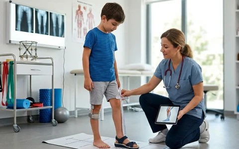
- Clinical measurement: blocks of different heights are placed under the shorter leg until the pelvis is level. Tape-measure readings are useful but not reliable enough on their own.
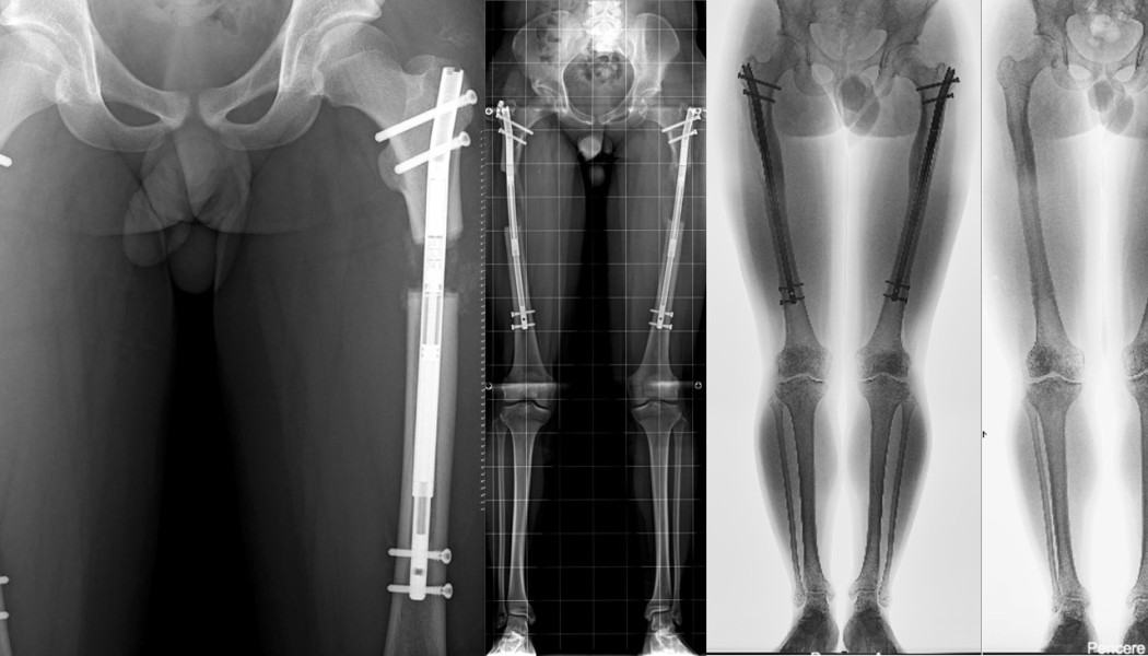
- Standing full-length leg X-ray: shows the whole leg from hip to ankle on a single image, revealing both the length difference and any associated deformity.
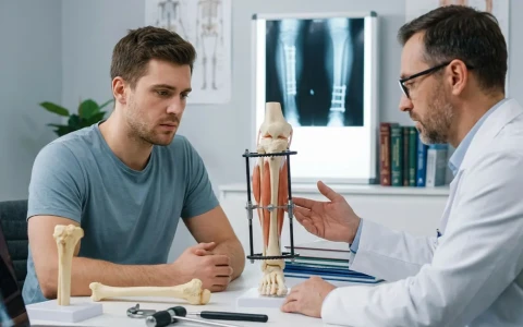
- Bone-by-bone measurement: establishes whether the difference comes from the thigh bone, the shin bone or both, which determines which bone is treated.
- CT or low-dose imaging: may be used for more accurate measurement, particularly when the knee cannot fully straighten.
- Bone age and growth prediction: in children, bone age is assessed from an X-ray of the hand and wrist, and the expected difference at the end of growth is calculated using methods such as the multiplier method or growth charts.
Joint stability is an equally important part of the assessment. In congenital shortening in particular, the hip and knee are checked to see whether they are ready for lengthening; if not, an operation to stabilise the joint may be planned first. Rotational problems and foot deformities are considered as part of the same plan.
Treatment options according to the size of the difference
The table below offers a general framework. The actual decision is made after examination and imaging, taking into account age, the cause of the difference, the state of the joints and the person's expectations.
| Method | Usually considered for | Advantages | Points to consider |
|---|---|---|---|
| Insole or shoe raise | Small differences; people who prefer not to have, or are unsuitable for, surgery | No operation; can be used straight away | Can be heavy and awkward for larger differences; does not remove the difference |
| Epiphysiodesis (stopping growth in the longer leg) | Growing children expected to have a moderate difference at the end of growth | Small incisions and a quick recovery | Performed on the healthy leg; timing depends on growth prediction; slightly reduces adult height |
| Shortening the longer leg | Selected adults with a moderate difference whose growth is complete | Equalisation in a single step | Performed on the healthy leg; may affect muscle strength; reduces height |
| Lengthening the shorter leg | Larger differences, associated deformities, skeletal dysplasias | Treats the short side; deformity can be corrected at the same time | Treatment lasts months; needs physiotherapy and close follow-up |
Very large and complex congenital differences may need more than one lengthening, spread across the growing years. In some rare situations, options based on a prosthesis (artificial limb) are also discussed openly with the family.
Epiphysiodesis or lengthening?
How is epiphysiodesis done?
Epiphysiodesis stops growth in the longer leg by closing its growth plate, usually around the knee, through small incisions. As the shorter leg continues to grow, the difference closes by the end of growth. The procedure is short, and children usually return to normal activities within a few weeks. The effect, however, is irreversible and depends entirely on timing: if it is done too early the difference may reverse, and if it is done too late the correction will be incomplete. Bone age and growth prediction are therefore assessed carefully, and the child is followed until growth has finished. Our article on leg length discrepancy in children gives more information on the options at this age.
How is medical limb lengthening done?
Lengthening relies on a biological principle called distraction osteogenesis. The bone is divided in a controlled way (an osteotomy) and, after a waiting period of a few days, the gap is opened by about 1 mm a day in small steps, while the body fills it with new bone (the regenerate). Once lengthening is complete, this new bone is allowed to harden (consolidation). Lengthening can be carried out with:
- External fixators: Ilizarov-type circular frames or monolateral fixators, which also allow deformities to be corrected at the same time as lengthening. Our page on Ilizarov and limb reconstruction surgery explains these in detail.
- Lengthening over a nail (LON): lengthening is done with an external fixator, after which a nail already placed inside the bone is locked, so the fixator can be removed earlier.
- Magnetic lengthening nails: nails placed inside the bone and lengthened from outside with a magnetic device, so no external frame is needed. The bone diameter, the state of the growth plates and the type of deformity must all be suitable.
Bone generally heals faster in children, but the need to protect growth plates and the size of the bones mean that not every method can be used at every age. The choice depends on the size and shape of the bone, any associated deformity and the state of the joints.
Limb lengthening in skeletal dysplasias and achondroplasia
In achondroplasia and similar skeletal dysplasias, lengthening may be considered to improve independence in daily life and to correct associated leg deformities. It is usually planned as a staged programme on both sides, involving the thigh and shin bones. In some patients, lengthening of the upper arm (humerus) is also considered to make personal care and everyday tasks easier; this is covered on our page on upper limb lengthening and deformity correction.
This is an extensive and lengthy process, so the decision should be made with the family, and wherever possible with the child, taking into account the child's age, expectations and psychosocial circumstances. New medicines that aim to support growth in children whose growth plates are still open have also been introduced in recent years; these are assessed together with paediatric endocrinologists and geneticists and can be considered alongside any surgical plan. Other problems specific to achondroplasia, such as narrowing of the spinal canal, are also part of the follow-up plan.
The lengthening process step by step
- Planning: the length difference, deformities, rotational alignment and joint stability are measured, and the amount and method of lengthening are decided.
- Surgery: the bone is divided in a controlled way and the chosen device is fitted; deformity can be corrected during the same operation if needed.
- Latency period: usually 5–7 days, allowing the first healing tissue to form.
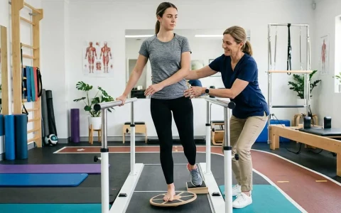
- Distraction phase: lengthening of about 1 mm a day, in several small increments. Regular X-rays and daily physiotherapy are essential at this stage to protect joint movement and muscle flexibility.
- Consolidation phase: partial weight-bearing continues until the new bone has hardened, which usually takes longer than the lengthening itself.
- Device removal and rebuilding strength: external fixators are removed once the bone is solid, and internal nails can usually be taken out in a planned operation after a year or two. A strengthening programme then continues.
The overall duration depends on the amount of lengthening, age and how quickly the bone heals, and it varies from person to person; it is usually longer in adults than in children. Planning school or work arrangements, getting around at home and the physiotherapy schedule in advance makes the process easier.
Possible risks and complications
Limb lengthening is a treatment that needs careful planning and close monitoring. The main problems that can arise are:
- Pin or wire site infection: common with external fixators; most cases settle with regular care and a short course of treatment.
- Joint stiffness and muscle tightness: muscles and tendons may struggle to keep pace with the lengthening bone, which can cause reduced knee bend or a foot that points downwards (equinus).
- Joint subluxation: particularly in congenital shortening, the hip or knee may partly dislocate, which is why joints are assessed in detail before lengthening.
- Premature or delayed consolidation: the new bone may harden too early or too slowly, requiring the rate of lengthening to be adjusted or a further procedure.
- Nerve stretch: if numbness or weakness develops, lengthening is slowed or paused.
- Other problems: drift of the alignment, device malfunction, and fracture of the new bone after the device has been removed.
Most complications can be managed if they are recognised early, which is why attending follow-up regularly and keeping up with physiotherapy are so important. Our article on complications in limb lengthening surgery provides more detail.
When should you seek advice?
An assessment by an orthopaedic surgeon is advisable in the following situations:
- A child who limps, walks on the toes of one foot or whose trouser legs hang noticeably unevenly
- A child who has had a fracture or bone infection and, months after healing, develops shortening or bowing of the leg
- A baby born with a limb difference, for an initial assessment and a monitoring plan
- A child with achondroplasia who has bow legs or whose short stature makes daily life difficult
- An adult with shortening, a limp and back or hip pain after a fracture
Early assessment is particularly important in growing children, because some options, such as epiphysiodesis, can only be used within a certain age range.
Prof. Dr. Halil İbrahim Balcı's approach
Prof. Dr. Halil İbrahim Balcı is a faculty member in the Department of Orthopaedics and Traumatology at Istanbul University's Istanbul Faculty of Medicine. At the Paley Institute in the USA he worked as a research fellow (2014) and observer (2019) in limb lengthening, deformity surgery and congenital bone anomalies. His presentations on four-segment lower limb lengthening and bilateral humeral lengthening in children with achondroplasia received awards at national and international congresses, and he is a co-editor of a Springer book on external fixator applications according to Ilizarov principles. Treatment plans are based on measurements, growth prediction and goals agreed with the family. Consultations are available in Turkish, English and French, and you can request one through the appointment page.
Key points
- Treatment of leg length discrepancy depends on the size and cause of the difference, age and any associated deformity.
- In children, predicting the difference at the end of growth is the basis of the treatment plan.
- Insoles for small differences, well-timed epiphysiodesis in growing children and lengthening of the shorter leg for larger differences are the main options.
- In skeletal dysplasias such as achondroplasia, lengthening is a long, staged process on both sides, decided together with the family.
- Close follow-up and regular physiotherapy during lengthening allow complications to be recognised early.