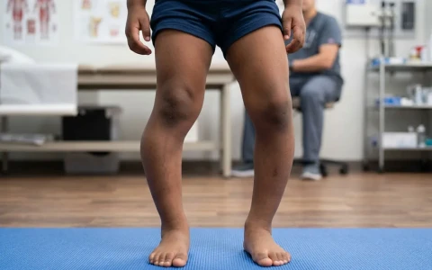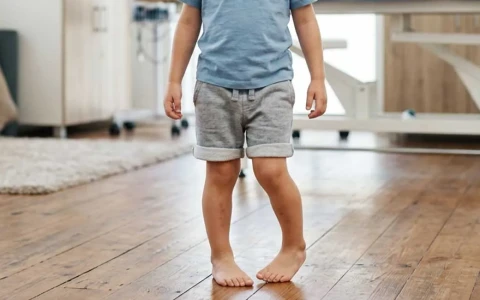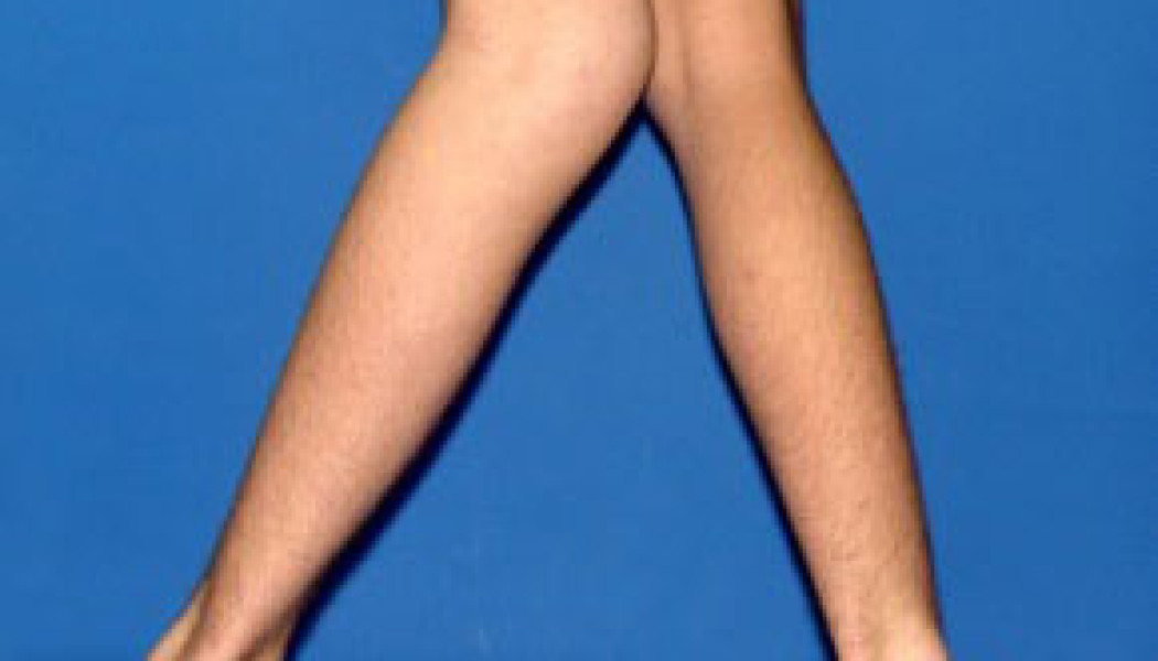
Leg bowing in children is, in most cases, a normal part of development: babies are born bow-legged, the knees drift towards knock knees in the pre-school years, and in most children the legs settle into adult alignment by around 7–8 years of age. In-toeing, out-toeing and flexible flat feet are also common in young children and usually improve on their own with growth. In a smaller number of children, however, a deformity is the sign of an underlying problem such as Blount disease, rickets, growth plate damage or a skeletal dysplasia, and needs treatment.
This page aims to help parents tell physiological (normal) alignment from pathological alignment caused by a condition, and to explain when watching and waiting is enough and when an assessment is needed. Rare conditions such as congenital limb deficiencies are covered on our page on rare childhood limb deformities, and leg deformity in people who have finished growing on our page on adult leg deformity correction.
When is leg bowing in children normal?
Knee alignment changes in a predictable sequence through childhood. The table below summarises this normal pattern; the age ranges can vary by a few months from child to child.
| Age | Expected alignment | What parents see |
|---|---|---|
| Birth to 18 months | Physiological bow legs (genu varum) | A gap between the knees when the ankles touch; may be obvious when walking starts |
| 18–24 months | Legs straighten | The bowed look fades |
| 3–4 years | Physiological knock knees (genu valgum) at their most marked | A gap between the ankles when the knees touch |
| 7–8 years | Close to adult alignment | Slight knock-knee position, similar to an adult |
Throughout this process, the alignment is similar on both sides, gradually improves, is not painful and the child's height is normal for their age. Physiological bowing does not need braces, special shoes or insoles.
Bow legs and knock knees: normal or not?
The question parents ask most often is whether their child's legs are normal. The features below help tell the difference:
| Feature | Physiological alignment | Alignment that needs assessment |
|---|---|---|
| Sides | Similar in both legs | One leg only, or clearly asymmetrical |
| Course | Improves with time | Gets worse, or fails to improve at the expected age |
| Age | Bow legs up to about 2 years, knock knees up to 7–8 years | Bow legs persisting beyond 2–3 years, marked knock knees after 8 years |
| Height | Normal for age | Clearly shorter than other children of the same age |
| Walking | No obvious problem | Knee lurching sideways when walking, limping, pain |
| History | Nothing notable | Previous fracture or infection, family history of a skeletal condition, nutritional problems |
For more on how bow legs and knock knees are managed in children, see our article on treating bowlegs and knock knees in children.
In-toeing and out-toeing: tibial torsion and femoral anteversion
When a child walks with the feet turned in (in-toeing) or out (out-toeing), the cause is usually the natural twist of the bones along their own axis. The likely cause can often be predicted from the child's age:
- Metatarsus adductus (infancy): the front of the foot curves inwards. Most flexible cases resolve on their own in the first years; stiffer ones may need stretching or casting.
- Internal tibial torsion (ages 1–4): an inward twist of the shin bone. It is the commonest cause of in-toeing in toddlers, and most cases resolve by school age. Our article on tibial torsion and in-toeing in children explains this in more detail.
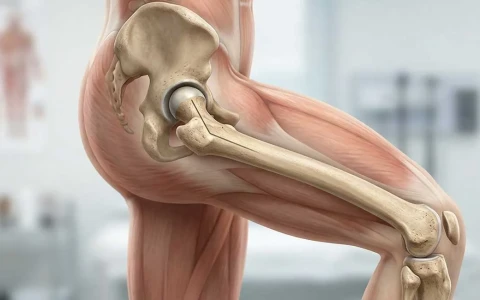
- Increased femoral anteversion (ages 3–8): the neck of the thigh bone is twisted further forwards than usual. These children's kneecaps point inwards, their legs swing outwards when they run, and they find sitting in a "W" position comfortable. W-sitting is usually a result of this anatomy rather than its cause. In most children it improves considerably with growth; see our article on femoral anteversion.
- External tibial torsion and out-toeing: an outward twist of the shin bone. Unlike inward twist, it can increase with age and in some children causes pain around the kneecap.
An important warning: new out-toeing, a limp and knee or groin pain in an overweight adolescent can be signs of a hip condition such as slipped capital femoral epiphysis, which needs urgent assessment. Knee pain does not always come from the knee.
Night splints, special shoes, insoles and bars that link the legs together do not change the natural course of rotational differences. Surgery (a derotation osteotomy) is considered only for severe twists that have not improved with growth and cause real functional problems, usually after the age of 8–10.
Causes of pathological bowing: Blount disease, rickets and others
Blount disease
Blount disease is a progressive bow-leg deformity caused by poor growth of the inner part of the growth plate at the top of the shin bone. The early-onset (infantile) form usually becomes obvious between the ages of 2 and 4; early walking and excess weight are among the risk factors. The adolescent form is seen mainly in overweight teenagers. It is distinguished from physiological bowing by measuring angles at the top of the tibia on standing X-rays. A brace may be tried in some young children at an early stage; in progressive cases, guided growth or a corrective osteotomy of the tibia may be needed, and in advanced stages and in adolescents, gradual correction with an external fixator. Because the deformity can recur despite treatment, follow-up until the end of growth is important. See our article on Blount disease for more.
Rickets
Rickets means that growing bone is not adequately mineralised. The commonest cause is a lack of vitamin D and calcium (nutritional rickets). In Turkey its frequency has fallen thanks to the national vitamin D programme for infants, but it is still seen. Bow legs or knock knees, widened wrists, bead-like swellings along the ribs, late walking and dental problems are warning signs. Some children have genetic forms of rickets involving phosphate loss, which do not respond to vitamin D alone and are managed together with paediatric endocrinologists. In rickets, the metabolic problem is treated first, and in young children much of the bowing can improve with that treatment alone. Guided growth or osteotomy is considered for deformity that persists once the metabolic problem is under control.
Other causes
- Growth plate damage: if part of a growth plate closes after a fracture or infection, the bone cannot grow on that side, and an increasing angulation develops.
- Knock knee after a fracture at the top of the tibia: in young children, a knock-knee deformity may appear in the months after this fracture; because it often corrects itself over the following years, it is usually monitored first.
- Skeletal dysplasias: achondroplasia and other dysplasias can cause bow legs or joint problems.
- Multiple hereditary exostoses: can lead to knock knees and an outward tilt at the ankle.
- Excess weight in adolescence: increases the load on the knees and can make knock knees more pronounced.
- Bone disorders related to kidney disease.
Flat feet (pes planus): when are they a problem?
Most young children's feet look flat. At this age, the fat pad in the sole and the flexibility of the ligaments hide the arch, which usually develops gradually over the first ten years. In a flexible flat foot, the arch appears and the heel turns inwards when the child stands on tiptoe; if there is no pain, no treatment is needed. Insoles do not build a lasting arch, although they can ease symptoms in children who have pain.
Flat feet should be assessed if there is:
- a stiff (rigid) flat foot, where no arch forms on tiptoe
- recurrent pain in the foot, leg or ankle, or frequent ankle sprains
- a tight Achilles tendon, with the heel not reaching the floor properly
- a flat foot on one side only, or one that appears in adolescence (possibly a congenital fusion between foot bones)
- in a newborn, a sole that is convex like the base of a rocking chair
Assessment: examination and imaging
Most leg alignment problems in children can be assessed by examination alone. The examination looks at height and weight, the distance between the knees and between the ankles when standing, walking pattern, joint flexibility and the rotational profile. The rotational profile measures the angle of the foot in relation to the direction of walking, hip rotation lying face down and the thigh–foot angle.
- Standing long-leg X-ray: not needed for typical physiological alignment; taken when there is asymmetry, progression, short stature or another concerning finding, which spares children unnecessary radiation.
- Blood tests: calcium, phosphate, alkaline phosphatase, vitamin D and parathyroid hormone levels when rickets is suspected.
- Hand and wrist X-ray (bone age): used to estimate remaining growth when guided growth is being planned.
- CT or MRI: needed only when surgery is planned for a rotational problem, or when growth plate damage is suspected.
Treatment options: monitoring, guided growth and osteotomy
For most children with leg alignment differences, treatment means monitoring with examinations, photographs and measurements at regular intervals. If there is an underlying condition, it is treated first. Physiotherapy supports strength and walking but does not change the shape of the bone, and special shoes and insoles do not correct bowing. When surgery is needed, there are two main approaches:
Guided growth
A small plate (often called an eight-plate) or a screw is placed through a small incision across the growth plate on the convex (longer) side of the curve. It temporarily slows growth on that side, and as the other side keeps growing the bone gradually straightens. For this to work, the child must have enough growth remaining. Correction takes place over months, with check-ups every few months, and the implant is removed once the leg is straight. If it is not removed in time, the leg can overcorrect in the opposite direction; after removal, some of the deformity can return, particularly in younger children.
Corrective osteotomy
For children who are close to or at the end of growth, for severe deformity, or when a growth plate is damaged, the bone is cut at a planned level and corrected. The correction can be completed during surgery and held with a plate or wires, or achieved gradually with an external fixator; if there is also shortening, lengthening can be included in the same treatment.
| Feature | Guided growth | Corrective osteotomy |
|---|---|---|
| Suitable child | A child with enough growth remaining | Near or at the end of growth, or with a severe or complex deformity |
| Procedure | Small incisions, usually a short hospital stay | The bone is cut and fixed |
| Correction | Over months, with growth | In theatre, or over weeks with a frame |
| Daily life | Quick return to normal activities | A period on crutches or in a frame until the bone heals |
| Follow-up | Regular check-ups and timely implant removal are essential | Regular check-ups until healing and the end of growth |
Possible risks and long-term follow-up
Surgery for leg alignment in children is planned, but like any operation it carries risks:
- Guided growth: loosening or breakage of the implant, overcorrection if follow-up visits are missed, partial return of the deformity after removal, and, rarely, early closure of the growth plate.
- Osteotomy: infection, slow bone healing, loss of correction, nerve or blood vessel problems (particularly the nerve that runs around the outer side of the knee when the top of the tibia is corrected) and excessive swelling of the leg.
- Recurrence: in Blount disease, rickets and skeletal dysplasias, the deformity can return with growth, which is why follow-up until the end of growth matters.
Whatever the treatment, regular check-ups while a child is still growing are key to maintaining the result. Outcomes and timelines vary from child to child.
When should you seek an assessment?
It is advisable to have your child assessed by an orthopaedic and trauma surgeon if you notice any of the following:
- bowing in one leg only, or clearly asymmetrical legs
- bow legs that persist or increase after the age of 2–3
- knock knees that are marked or increasing after the age of 8
- leg bowing together with clearly short stature for age
- a knee that lurches sideways when walking, a limp, pain or frequent falls
- bowing together with widened wrists, late walking or nutritional problems
- in-toeing or out-toeing that has not improved with growth and affects function
- painful or stiff flat feet
- a new limp, out-toeing and knee or groin pain in an overweight adolescent, which should be assessed without delay
For other topics in children's bone and joint care, see our paediatric orthopaedics page.
Prof. Dr. Balcı's approach to leg alignment problems in children
Prof. Dr. Halil İbrahim Balcı is a faculty member in the Department of Orthopaedics and Traumatology at Istanbul University, Istanbul Faculty of Medicine. At the Paley Institute in the United States he worked as a research fellow and later as an observer in paediatric orthopaedics, deformity surgery and Ilizarov techniques, and he has contributed to more than 50 international publications in paediatric orthopaedics and deformity. With leg bowing in children, his priority is to spare normal developmental alignment from unnecessary treatment while recognising, in good time, the children who genuinely need it; decisions are made with the family after examination and, where needed, imaging. Consultations are available in Turkish, English and French through our appointment page.
Key points
- Bow legs in infancy and knock knees at 3–4 years are normal; most children reach adult alignment by around 7–8 years.
- Bowing that is one-sided, progressive, painful or associated with short stature should be assessed.
- In-toeing and flexible flat feet usually improve with growth; special shoes and insoles do not change their natural course.
- Blount disease, rickets and growth plate damage are the main causes that need treatment.
- Guided growth and osteotomy are effective options for suitable children; outcomes vary from child to child.