Lower limb rotational deformities are orthopedic conditions characterized by excessive rotation of the femur (thigh bone) or tibia (shin bone) around their own axes beyond anatomical norms. This condition can directly affect patients’ gait patterns (such as in-toeing or out-toeing), joint mechanics, and overall quality of life. Although often identified during childhood, symptoms may persist or manifest in adulthood. Without accurate diagnosis, these deformities can lead to early degeneration of the hip and knee joints.
In this article, we will examine lower limb rotational deformities starting from anatomical fundamentals, progressing through current diagnostic methods and surgical treatment options. Our goal is to provide patients and caregivers with reliable, scientifically based information about this complex process.
Introduction to Lower Limb Rotational Deformities
Rotational deformities are often less noticeable than frontal plane deformities (like bowlegs or knock-knees), but their biomechanical impact can be significant. The alignment of the lower limb extends from the hip joint to the ankle via a complex mechanical axis. Any rotational deviation along this axis disrupts load distribution and increases joint stress.
Clinically, patients commonly present with complaints such as “feet crossing while walking,” “frequent falls during running,” or “knee pain.” Identifying the underlying rotational abnormality is the first and critical step towards successful treatment.
Anatomical and Biomechanical Foundations
The rotational profile of the lower limb is determined by the angular relationship of the femur and tibia with each other and the pelvis. Normal gait requires these bones to maintain specific rotational angles. Natural changes in these angles occur from birth to adulthood, but values outside certain limits are pathologic.
Femoral Anteversion and Tibial Torsion
The two most common components of rotational deformities are femoral anteversion and tibial torsion. Femoral anteversion refers to the forward rotation of the femoral neck relative to the knee axis. Increased femoral anteversion often causes the patient to walk with inwardly turned feet (in-toeing). Tibial torsion is the rotation of the tibia along its own axis, which can be internal or external.
Causes of Rotational Deformities
Rotational deformities may arise from congenital, developmental, or acquired (traumatic) causes. Congenital cases may involve intrauterine positioning or genetic factors, while developmental causes often include asymmetric growth of the growth plates.
Post-traumatic deformities generally result from malunion of fractures. Childhood fractures or growth plate injuries can cause rotational abnormalities as the bone grows. Additionally, neuromuscular conditions such as cerebral palsy may lead to secondary rotational deformities due to muscle imbalance.
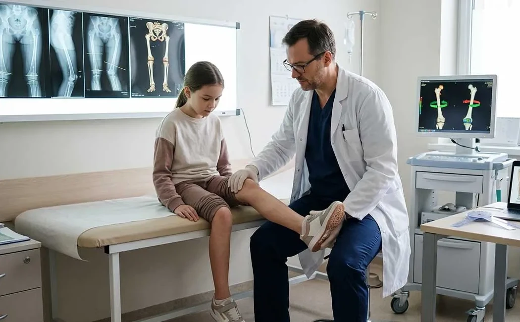
Diagnostic Methods
Diagnosis begins with a meticulous clinical examination. The physician analyses gait (gait analysis) and performs a specific set of tests known as the “rotational profile,” assessing parameters such as hip rotation range, thigh-foot angle, and heel bisector angle.
To confirm clinical suspicion and plan surgery, radiological imaging is indispensable. While standard X-rays reveal bone structures, computed tomography (CT) remains the most reliable method for accurate measurement of rotational angles. CT allows millimeter-level assessment of femoral and tibial torsion angles to determine deviations from normal values.
Treatment Methods
The treatment strategy depends on deformity severity, patient age, symptomatology, and underlying cause. Mild deformities without functional impairment may be monitored, whereas painful or gait-disrupting deformities require active intervention. Surgical decision-making considers the overall anatomical context in line with fundamental principles of lower limb deformity surgery.
Non-Surgical Options
Some mild rotational abnormalities in childhood may spontaneously improve with growth (remodelling). During this period, patient education and regular follow-up are important. Although orthotic devices and special shoes are sometimes considered, their effectiveness in correcting bony rotation is debated in scientific literature. Physical therapy and muscle strengthening exercises serve as supportive treatments to manage muscle imbalances and improve gait quality.
Surgical Intervention: Derotation Osteotomy
The primary treatment for fixed and severe rotational deformities is derotation osteotomy. This procedure involves controlled cutting of the bone (osteotomy), realigning it to the correct angle, and fixation with plates and screws or intramedullary nails.
In some complex cases, limb length discrepancies can be corrected simultaneously. Combined approaches using bone lengthening techniques may be considered. Preservation of joint health and delay of prosthetic surgery are also important considerations, with strategies involving joint-preserving surgeries in rotational deformity management.
Approach in Pediatric Patients
Rotational issues in children require a distinct approach from adults. Open growth plates provide both advantages (potential for spontaneous correction) and risks (risk of growth plate injury). Pediatric lower limb deformities are often within physiological developmental limits and tend to resolve over time. Nonetheless, unilateral, progressive, or painful deformities warrant thorough evaluation. Treatment timing should be carefully planned according to the child's age and natural progression of the deformity.
Complications and Follow-Up
As with any surgery, rotational deformity correction carries risks such as infection, nerve injury, or delayed union/nonunion. Treating deformities caused by malunited fractures may be complicated by existing hardware or soft tissue adhesions.
Delayed healing requires specific nonunion surgical protocols. Untreated or late-treated severe deformities may lead to long-term joint arthritis. At this stage, options such as hip replacement and advanced deformity management should be discussed. Modern medicine also includes biological treatment methods to support healing in nonunion fractures.
Conclusion and Informational Note
Lower limb rotational deformities require accurate diagnosis and expert management, as untreated cases can significantly decrease quality of life. Surgical methods aimed at restoring anatomy and function are applied successfully today. However, treatment must be individualized. This article is for informational purposes only; consultation with a specialist is essential for accurate diagnosis and appropriate treatment planning.
All information provided is for general informational purposes only and does not constitute medical advice. The content is not intended to replace individual diagnosis, treatment, or professional consultation. Diagnosis and treatment decisions should only be made following an in-person examination by a qualified healthcare provider. Since each patient's clinical condition is unique, the surgical or non-surgical methods applied may vary accordingly. The information is based on current scientific knowledge and up-to-date medical practices. Do not delay seeking professional medical care based on the information provided here.