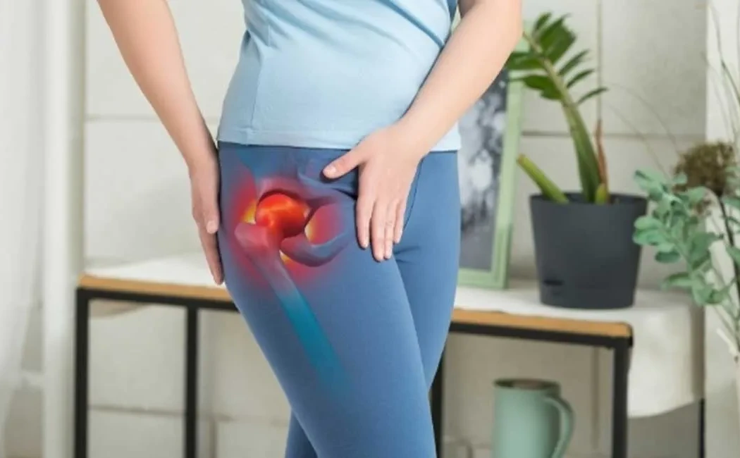
Hip impingement syndrome (Femoroacetabular Impingement - FAI) occurs due to abnormal contact between the bony structures forming the hip joint. This abnormal contact can lead to damage of the joint surfaces, pain, and limited movement. It is especially common among young, middle-aged, and active individuals, particularly athletes. If left untreated, it may contribute to the development of hip osteoarthritis.
Anatomically, the hip joint is formed by the combination of the femoral head and the acetabulum (the socket of the hip bone). In impingement syndrome, there is abnormal friction and contact between these two structures beyond normal limits. FAI is typically classified into three types: femoral, acetabular, and mixed. The femoral type involves a deformity in the neck region of the femoral head. The acetabular type is characterized by a deeper or protruding rim of the hip socket. The mixed type includes deformities in both structures.
Early symptoms often begin with either no symptoms or mild pain, making patient awareness crucial. The most common clinical signs include deep hip pain during walking or standing up from a seated position, tenderness on the front or side of the thigh, restricted hip movement, and a catching sensation during motion. Pain is usually felt in the groin area and intensifies particularly with flexion (lifting the leg forward) movements. Symptoms worsen after prolonged sitting or walking. If these signs are neglected, damage to the cartilage and labrum (the ring of cartilage around the joint) progresses, leading to functional loss of the hip joint.
Diagnosis relies on detailed patient history, clinical evaluation, and physical examination. The impingement test applied to the hip joint provokes pain. Definitive diagnosis requires imaging techniques. X-rays assess bone abnormalities such as femoral head deformities or acetabular protrusions. Magnetic resonance imaging (MRI) reveals damage to soft tissues, cartilage, and the labrum. These imaging methods are critical for early diagnosis and planning appropriate treatment.
Treatment varies according to patient age, severity of symptoms, and degree of joint damage. Conservative treatments initially include tailored exercise programs and physical therapy aimed at strengthening muscles, increasing joint range of motion, and reducing pain. Medications such as analgesics and anti-inflammatory drugs may support symptom management.
Surgical treatment is considered if conservative methods fail or if there is advanced structural deformity. Arthroscopic surgery is the most successful approach in this area. Minimally invasive arthroscopic hip surgery allows correction of deformities and repair of cartilage and the labrum. Postoperative rehabilitation is crucial for recovery, starting with physiotherapy to regain motion and progressively increasing strength.
Early identification and treatment of hip impingement syndrome can preserve long-term hip function in most patients and reduce the risk of osteoarthritis development. Hence, individuals experiencing hip pain and movement limitations are advised to consult an orthopedic specialist promptly.