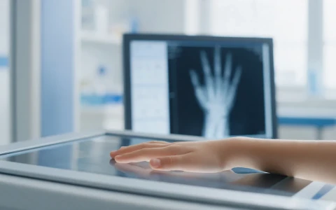
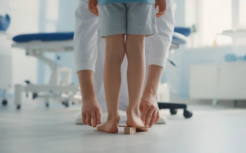
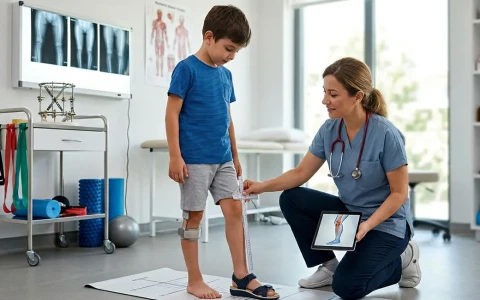
Leg length difference and alignment are measured in detail on a standing full-length leg X-ray. We explain the long-leg X-ray and other methods, how to prepare and how the results are interpreted.
On this page 8 sections
- Why does leg length difference need accurate measurement?
- Clinical measurement: tape measure and block test
- What is a leg length X-ray and how is it taken?
- Other methods: CT scanogram and EOS
- What does the report look at?
- Measurement and growth prediction in children
- When should you see a doctor?
- Key points
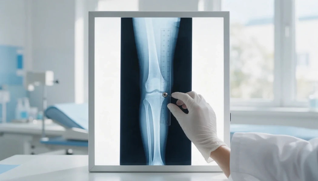
A leg length X-ray is a calibrated, standing radiograph that shows the whole of both legs, from the hips to the ankles, so that a leg length difference and any bowing can be measured accurately. You may also hear it called a long-leg X-ray, a full-length standing film, an orthoroentgenogram or a teleoroentgenogram. A tape measure gives a useful estimate, but this X-ray is the key test because it measures the thigh and shin bones separately, to the millimetre, together with the joint angles.
This guide explains the different ways leg length difference is measured, how a long-leg X-ray compares with other imaging, how to prepare and what the report looks at. Treatment decisions are always made by combining the measurements with the findings of a clinical examination.
Why does leg length difference need accurate measurement?
Many people have a difference of a few millimetres between their legs, and it usually causes no problems. As the difference grows, however, it can cause limping, a tilted pelvis and strain on the back, hips and knees. The body tries to compensate, for example by walking on tiptoe on the short side, bending the knee on the long side or curving the spine.
Not every leg that looks short is actually shorter. Stiffness of the hip or knee, a tilted pelvis or a curved spine can make a leg appear short even when the bones are equal; this is called an apparent (functional) leg length difference. The true amount, which bone it comes from and, in children, how it will change with growth all shape the treatment options. These are discussed on our page on limb lengthening for leg length discrepancy.
Clinical measurement: tape measure and block test
The first step at examination is usually a tape measure. The distance from the bony point at the front of the pelvis to the inner ankle bone reflects true leg length, while the distance from the navel to the inner ankle reflects any apparent difference. It is quick and involves no radiation, but finding bony landmarks through soft tissue can introduce errors of a centimetre or so.
In the block test, you stand while blocks of known thickness are placed under the shorter leg until the pelvis is level. This shows what the difference means in everyday life and helps when planning a shoe raise or insole. Neither method shows whether the difference lies in the thigh or the shin, so both are complementary rather than definitive.
What is a leg length X-ray and how is it taken?
For a leg length X-ray you stand, and both legs from the hips to the ankles are captured in one long image. Modern digital systems take several overlapping images and stitch them together automatically. A calibration marker, such as a metal ball of known diameter or a ruler, is included so that the magnification inherent in X-rays can be corrected and true lengths calculated.
The classic orthoroentgenogram combined three separate exposures, over the hip, knee and ankle, with a ruler on one long film, while a teleoroentgenogram is a single long exposure taken from a greater distance. Today the terms are often used interchangeably, and all share the same aim: a measurable image of the whole leg, standing and bearing weight. A side view is sometimes added to assess bowing from front to back.
How should you prepare?
- Wear clothes without metal buttons, zips, belts or jewellery; a gown may be provided.
- Shoes, insoles and orthoses are usually removed unless you are told otherwise.
- Always tell the radiographer if there is any chance you could be pregnant.
- Mention any implants or external fixator, if you cannot fully straighten a knee, or if you normally use a shoe raise.
- Stand with your knees as straight as possible, kneecaps facing forwards and weight spread evenly on both legs.
- If the difference is marked, a block may be placed under the shorter leg to level the pelvis, as your doctor requests; this is noted on the report.
- Bring previous X-rays; for follow-up, using the same technique each time makes comparison easier.
Other methods: CT scanogram and EOS
| Method | How it is done | Advantages | Limitations |
|---|---|---|---|
| Tape measure | Measured between bony landmarks at examination | Quick, no radiation | Larger error; cannot separate the bones |
| Block test | Blocks under the shorter leg while standing | Shows the functional difference | No bone-level measurement |
| Standing full-leg X-ray | Calibrated X-ray from hips to ankles | Length, alignment and joint angles under load | Posture errors can affect results |
| CT scanogram | Low-dose CT lying down | Precise lengths; rotation can be measured too | Not weight-bearing; limited alignment assessment |
| EOS imaging | Standing, front and side images taken together | Low dose, three-dimensional measurements | Not available everywhere |
A CT scanogram can make length measurement easier for people who cannot fully straighten a knee. If a rotational problem is suspected, the twist of the thigh and shin bones can also be measured with CT or MRI. Your doctor will choose the method according to the clinical question and local facilities.
What does the report look at?
A long-leg X-ray answers much more than "how many centimetres?". The assessment usually covers:
- Bone lengths: the length of the thigh and shin bones on each side, and the total difference
- Mechanical axis: where a line from the centre of the hip to the centre of the ankle crosses the knee; a shift towards the inner side suggests bow legs, towards the outer side knock knees
- Joint angles: angles at the lower end of the femur and upper end of the tibia (normally about 85–90 degrees) show which bone the deformity comes from
- Apex of the deformity: if corrective surgery is planned, where and how much the bone is angled
- Growth plates: in children, whether they are still open
Some factors can affect the results. A knee that does not fully straighten, kneecaps pointing inwards or outwards rather than forwards, and a tilted pelvis can all alter length and angle measurements. Differences of a few millimetres between images taken at different centres or in different positions are therefore common, and during follow-up the trend across images taken with the same technique matters more than any single measurement.
Our article on CORA and the principles of deformity correction explains how the apex is found. These measurements also form the basis of planning for leg deformity correction.
Measurement and growth prediction in children
In children, leg length difference can change with growth. Differences caused by congenital conditions usually increase as the child grows, while some that follow fractures stay the same or lessen. Long-leg X-rays are therefore often repeated every 6 to 12 months during growth, and a hand and wrist X-ray may be used to estimate bone age.
The measurements are combined with prediction methods, such as the Paley multiplier method, to estimate the difference at the end of growth. This helps time procedures that guide growth or decide whether lengthening may be needed later. Broadly, small differences are often managed with observation or a shoe raise, moderate ones in growing children with procedures that balance growth, and larger ones with bone lengthening, although each decision is individual. See our article on leg length discrepancy in children for more.
Radiation doses from modern digital equipment are low; even so, the principle is to avoid unnecessary repeats and to image only at intervals that will influence treatment decisions.
When should you see a doctor?
- A limp, or leaning onto one side when walking
- One shoe wearing out faster, or trouser legs looking uneven
- Back, hip or knee pain together with a leg length difference
- A child walking on tiptoe on one side, or an asymmetry that becomes more obvious over time
- A change in leg length or shape after a childhood fracture
- Marked bow legs or knock knees
Measurements should always be interpreted alongside the examination and the patient's age. Prof. Dr. Halil İbrahim Balcı's international publications focus mainly on deformity, limb lengthening and paediatric orthopaedics.
Key points
- A leg length X-ray is the core test for measuring leg length difference and alignment, standing and calibrated.
- Tape measures and block tests are useful but cannot show which bone the difference comes from.
- CT scanograms and EOS imaging are complementary or alternative options in suitable situations.
- Correct positioning, metal-free clothing and bringing previous images all improve measurement quality.
- In children, regular measurements help predict the final difference and time any treatment.
Frequently Asked Questions
A standard leg X-ray usually shows one bone or joint and has no scale. A leg length X-ray captures both legs from the hips to the ankles in a single image, taken standing and weight-bearing, with a calibration marker. This allows the thigh and shin bones to be measured separately and the leg's load axis and joint angles to be calculated. That information is central to planning treatment for leg length difference and bowing.
With modern digital equipment, the radiation dose for a full-length leg X-ray is low, and a single examination is not expected to pose a meaningful health risk. As with any X-ray, unnecessary repeats are avoided and images are taken only when they will influence treatment. Always mention any possibility of pregnancy. For children who need frequent follow-up, lower-dose systems such as EOS may be used where available.
It depends on the cause and size of the difference and on the child's age. For growing children whose difference is being monitored, measurements are often repeated every 6 to 12 months. Checks may be more frequent if the difference is increasing quickly or if a growth-guiding procedure is being planned, and less frequent if it is stable. The aim is to predict the difference at the end of growth accurately and to time treatment well. Your doctor will set the schedule.
A tape measure is a quick, radiation-free first step and is useful for spotting an obvious difference. Because bony landmarks have to be located through soft tissue, however, it can be out by a centimetre or so, and it cannot tell whether the difference lies in the thigh or the shin. If treatment is being considered, or a child's difference needs monitoring through growth, a calibrated image such as a long-leg X-ray is needed.
Many people have a difference of up to about a centimetre, and it often causes no symptoms. As the difference increases, limping and strain on the back, hips or knees become more likely. The number of centimetres alone does not decide treatment, though; symptoms, the cause, the person's age and, in children, the expected change with growth are all taken into account. That is why assessment combines examination with accurate measurement.
This article is for general information only and does not replace a medical examination, diagnosis or treatment. Treatment options and outcomes vary from person to person.
Related treatment area Adult Leg Deformity Correction: Bow Legs and Knock Knees