Distal femoral osteotomy realigns the lower thigh bone in knock knees to reduce overload on the outer part of the knee. We explain who may be suitable, how it is planned and what recovery involves.
On this page 8 sections
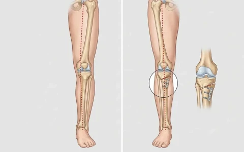
Distal femoral osteotomy (DFO) is a joint-preserving operation in which the lower end of the thigh bone (femur), just above the knee, is cut in a controlled way and its angle changed. It is most often used in young and active adults whose knock knees (genu valgum) overload the outer part of the knee, causing early arthritis or cartilage damage there. The aim is to rebalance the load-bearing axis of the leg, reduce pressure on the outer compartment and help the person keep their own knee joint for as long as possible.
You can think of DFO as the knock-knee counterpart of the high tibial osteotomy used for bow legs. Not every knock knee needs surgery; the decision is made individually after a standing full-leg X-ray, examination and, if needed, an MRI scan.
How do knock knees affect the knee?
In a normally aligned leg, the mechanical axis, a line from the centre of the hip to the centre of the ankle, passes close to the middle of the knee. In knock knees this line shifts towards the outer side. As a result, the cartilage and meniscus on the outer side carry more load, the ligament on the inner side is stretched and the kneecap may not track properly. Over the years this can cause pain and arthritis in the outer compartment.
Common causes of knock knees in adults include:
- Knock knees from childhood that did not correct themselves
- Fractures of the lower thigh bone or upper shin bone that healed crooked
- Extensive removal of the outer meniscus
- Previous rickets, some skeletal dysplasias and inflammatory joint disease
In adult knock knees the deformity usually lies in the lower end of the thigh bone, which is why correction is so often done there. Less often it comes from the shin bone or both bones, and planning changes accordingly. For an overview of knock knees in children and adults, see our article on genu valgum.
Who is distal femoral osteotomy suitable for?
DFO is usually considered for people who have:
- Knock knees confirmed on imaging, with the deformity arising from the lower thigh bone
- Pain on the outer side of the knee that worsens with activity, or early arthritis there
- Relatively healthy cartilage on the inner side of the knee and behind the kneecap
- Good knee movement and no major ligament instability
- A young or middle-aged, active lifestyle they wish to maintain
DFO may also be planned to protect cartilage repair or meniscal transplantation on the outer side of the knee, or for kneecap instability associated with knock knees. It is generally not a suitable option for advanced arthritis affecting all three compartments, inflammatory arthritis, marked stiffness or a knee that cannot straighten fully; in these situations knee replacement or other treatments are considered. Excess weight and smoking can also hinder bone healing.
Planning before surgery: which tests are needed?
Planning is based on a standing full-length leg X-ray. It shows how far the mechanical axis has moved to the outer side, and by measuring the joint angles of the lower femur and upper tibia it reveals which bone the deformity comes from. This analysis is a core principle of adult leg deformity correction.
Standing knee X-rays and kneecap views are also taken, an MRI may be used to assess cartilage, menisci and ligaments, and a CT scan may be requested if a rotational problem is suspected. The aim is to bring the load-bearing axis close to the centre of the knee. Overcorrection is avoided, because it would shift excess load onto the inner compartment instead, so the amount of correction is calculated degree by degree.
How is distal femoral osteotomy performed?
Opening-wedge technique
The bone is reached through an incision on the outer side of the thigh. It is cut from the outside inwards, leaving a thin bridge of bone (the hinge) on the inner side. The cut is opened by the planned amount and held with a locking plate, and the gap may be filled with bone graft or a bone substitute if needed.
Closing-wedge technique
Through the inner side of the thigh, a precisely calculated wedge of bone is removed and the bone ends are brought together and fixed with a plate. Because the bone surfaces are in direct contact, this can favour healing. The choice of technique depends on the size of the deformity, leg length, bone quality and the surgeon's plan.
For large deformities, those in more than one plane, or those combined with leg shortening, gradual correction with an external fixator over days to weeks is another option. X-ray guidance is used during the operation, and knee arthroscopy can be performed at the same time if needed.
| Feature | Distal femoral osteotomy | High tibial osteotomy |
|---|---|---|
| Bone operated on | Lower end of the thigh bone | Upper end of the shin bone |
| Typical alignment | Knock knees (valgus) | Bow legs (varus) |
| Compartment offloaded | Outer (lateral) side of the knee | Inner (medial) side of the knee |
| Usual fixation | Locking plate | Locking plate |
| Planning goal | Load axis close to the centre of the knee | Load axis slightly to the outer side of centre |
For the bow-leg equivalent, see our article on high tibial osteotomy.
Recovery after surgery
The hospital stay is usually a few days. Pain relief, ice and keeping the leg raised help in the first days. Knee movement exercises often start early, and measures to prevent blood clots follow your surgeon's advice.
For the first weeks you walk with crutches, putting partial weight through the leg, and weight-bearing is increased step by step as X-rays show healing. Bone union usually takes a few months. Desk work is often possible after a few weeks, while driving depends on which leg was operated on and how healing progresses. Returning to sport generally takes months, with physiotherapy playing a central role, and the full benefit may take close to a year to judge. Recovery varies from person to person.
Physiotherapy focuses on maintaining knee movement, rebuilding the strength of the quadriceps at the front of the thigh and restoring a normal walking pattern. When your surgeon agrees, low-impact activities such as static cycling and swimming are often the first to resume, while running and jumping sports are reintroduced gradually once bone healing and muscle strength allow.
Possible risks and the link with knee replacement
Like any operation, DFO carries risks:
- Delayed union or nonunion
- Fracture at the hinge or loss of correction
- Under- or overcorrection
- Infection, blood clots and knee stiffness
- Rarely, nerve or blood vessel injury
- Irritation from the plate and the possible need to remove it later
DFO does not reverse existing cartilage damage; its purpose is to relieve symptoms by reducing load and to slow further wear. Some people may still need a knee replacement in the future. A knee replacement after a previous osteotomy can require a little more planning but is usually feasible. For knock knees caused by a fracture that healed crooked, see our page on deformity and malunion surgery.
When should you see a doctor?
- Persistent pain on the outer side of the knee with walking, stairs or sport
- Knock knees that seem to be increasing over the years
- Recurrent swelling, a feeling of the knee giving way or the kneecap dislocating
- A new angulation of the leg after a fracture around the knee
- After surgery: fever, wound discharge, calf swelling or a sudden increase in pain
A sound decision requires detailed analysis of the deformity together with an assessment of the cartilage. Prof. Dr. Halil İbrahim Balcı worked as a research fellow in deformity surgery at the Paley Institute in the United States and has published internationally on lower limb deformities.
Key points
- Distal femoral osteotomy realigns the lower thigh bone in knock knees to reduce load on the outer knee.
- Suitable candidates are usually active adults with early outer-compartment damage and relatively healthy cartilage elsewhere.
- Planning relies on a standing full-leg X-ray and, if needed, MRI; overcorrection is avoided.
- Recovery starts with crutches and partial weight-bearing; bone healing takes a few months.
- DFO does not cure arthritis, but in suitable patients it aims to help preserve the natural knee for longer.
Frequently Asked Questions
Most patients are helped up on crutches by a physiotherapist soon after surgery. For the first weeks, partial weight is usually allowed through the leg, and as follow-up X-rays show healing, weight-bearing is increased gradually and the need for crutches falls. Walking unaided with full weight is often possible somewhere between a few weeks and a few months. The timescale depends on the technique used, bone quality and how healing progresses.
DFO does not reverse existing cartilage damage, so it cannot be said to prevent knee replacement for certain. Its aim, in suitable patients, is to relieve symptoms by reducing load on the outer compartment and to slow the progression of arthritis, which may postpone the need for a replacement. Some people will still need one later, and a knee replacement after a previous osteotomy is usually feasible.
Once the bone has healed, the plate can often be left in place. In some people, however, a plate on the side of the thigh irritates the overlying muscle and tissues, particularly when bending the knee. In that case removal can be considered once X-rays confirm full healing. If a knee replacement is planned in the future, the plate may be removed beforehand or during that operation. The decision is made after examination.
Both are joint-preserving operations that change the leg's alignment to reduce load on the damaged part of the knee. Distal femoral osteotomy is performed on the lower end of the thigh bone and usually offloads the outer side of the knee in knock knees. High tibial osteotomy is performed on the upper end of the shin bone and mostly offloads the inner side in bow legs. Which one is appropriate depends on where the deformity lies.
When both legs have knock knees, operating on both in one session is technically possible, but the operations are usually planned a few months apart. Having one sound leg to bear weight on during recovery makes daily life and rehabilitation much easier. In certain situations simultaneous surgery may be preferred. The decision is made together, taking into account the size of the deformity, general health and home circumstances.
This article is for general information only and does not replace a medical examination, diagnosis or treatment. Treatment options and outcomes vary from person to person.
Related treatment area Adult Leg Deformity Correction: Bow Legs and Knock Knees