A bone age X-ray shows how mature a child's skeleton is from an image of the left hand and wrist. We explain the Greulich-Pyle and Tanner-Whitehouse methods, what results mean and orthopaedic uses.

On this page 8 sections
- What is bone age and how does it differ from actual age?
- How is a bone age X-ray taken?
- How is bone age measured? Greulich-Pyle, Tanner-Whitehouse and other methods
- What does a delayed or advanced bone age mean?
- When is bone age used in orthopaedics?
- The limits of bone age: how should the result be interpreted?
- When should you see a doctor?
- Key points
Bone age is a measure of how mature a child's skeleton is, and it is usually worked out from a single X-ray of the left hand and wrist. On a bone age X-ray, the shape and size of the hand and wrist bones and the appearance of the growth plates are compared with standards prepared for each age and sex, and the result is reported as a value such as "bone age 11 years". A bone age clearly behind or ahead of a child's actual age helps to show what stage of growth the child has reached and how much growing time is left.
In paediatric endocrinology, bone age is used to assess short stature, tall stature and the timing of puberty; in orthopaedics, it helps to time treatment for leg length differences and leg deformities. This article explains in plain language how a bone age X-ray is taken, how it is assessed, what the result tells us and where its limits lie.
What is bone age and how does it differ from actual age?
A child's chronological age is simply the time since they were born. Bone age reflects how far the skeleton has matured. Children do not mature at the same pace: of two twelve-year-olds in the same class, one may enter puberty early and finish growing sooner, while the other carries on growing for several more years. Genetics, the timing of puberty, hormones, nutrition and general health all play a part.
It is common, and usually normal, for bone age to differ somewhat from chronological age; differences of a few months, and even of one to two years, can be seen in healthy children. Bone age on its own is therefore not a sign of illness. Its meaning becomes clear only when it is considered together with the child's height, growth rate, parents' heights and signs of puberty.
How is a bone age X-ray taken?
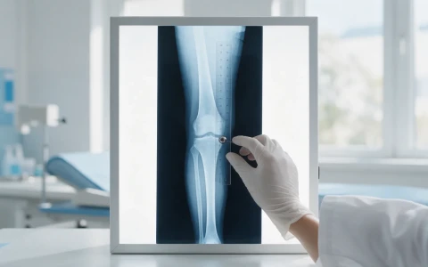
Bone age is usually assessed from a single front-view X-ray of the left hand and wrist. The child places their hand palm down on the X-ray plate with the fingers slightly apart, and the exposure takes a few seconds. No fasting, medication or special preparation is needed; rings and bracelets are simply removed.
The left hand is used because the reference standards were developed from left-hand X-rays. In addition, most people are right-handed, so signs of past injury are less common in the left hand. The radiation dose from a bone age X-ray is very low, generally less than the dose received from natural background radiation in a single day.
In very young infants a knee or foot X-ray is sometimes used, and around the rapid growth spurt of early puberty an elbow X-ray may also be used for orthopaedic planning.
How is bone age measured? Greulich-Pyle, Tanner-Whitehouse and other methods
Once taken, the image is assessed by a radiologist or by a paediatric endocrinologist or orthopaedic surgeon. The most widely used methods are:
- The Greulich-Pyle atlas: the child's X-ray is compared with an atlas of example X-rays for each age and sex, and the age of the closest match is taken as the bone age. It is quick and widely used.
- The Tanner-Whitehouse method: instead of a single overall comparison, the ends of the forearm bones and each of the hand and finger bones are scored individually by maturity stage, and bone age is calculated from the total score. It is more detailed and takes longer.
- Computer-assisted software: analyses the image automatically and calculates a bone age based on these methods. It is increasingly common, although the results are still interpreted by a doctor.
- The elbow method: during the growth spurt at the start of puberty, changes in the hand bones can be less distinct, so assessments based on an elbow X-ray are also used, particularly in orthopaedics.
| Method | Strength | Limitation |
|---|---|---|
| Greulich-Pyle | Quick, widely used and easily available | Results can vary between assessors; based on an older reference population |
| Tanner-Whitehouse | Detailed, bone-by-bone assessment | Takes longer and requires experience |
| Computer-assisted | Reproducible and independent of the assessor | Depends on image quality; still needs a doctor's interpretation |
| Elbow method | Useful during the growth spurt of early puberty | Only applicable within a limited age range |
What does a delayed or advanced bone age mean?
A delayed bone age means the skeleton is less mature than expected for the child's actual age, which usually indicates a longer period of growth ahead. The most common cause is constitutional delay of growth and puberty, often described as being a "late developer", which frequently runs in families. Thyroid hormone or growth hormone deficiency, long-term chronic illness and undernutrition can also delay bone age.
An advanced bone age means the skeleton is more mature than expected and suggests that growth may finish earlier. Early puberty, excess weight and some hormonal conditions can advance bone age, and some children from tall families also have a slightly advanced bone age.
Bone age and height measurements can be combined to predict adult height, but these predictions carry a margin of error of several centimetres and need to be reviewed over time. Interpreting bone age in terms of height and puberty is the role of paediatricians and paediatric endocrinologists, and families are advised not to interpret results on their own.
When is bone age used in orthopaedics?
Many treatments in paediatric orthopaedics either harness growth or wait for it to finish, so knowing how much growth a child has left is the basis of good timing. Bone age is assessed particularly in the following situations:
- Leg length difference: predicting the difference expected at the end of growth relies on bone age, alongside repeated leg measurements and growth prediction methods.
- Timing of epiphysiodesis: stopping the growth plate in the longer leg has to be timed so that the shorter leg catches up exactly as growth ends.
- Guided growth (8-plates): enough growth must remain for knock knees or bow legs to correct with growth.
- Timing of deformity correction: it helps decide whether a deformity should be treated with small procedures that use growth, or with an osteotomy once growth has finished.
- Scoliosis follow-up: spinal curves are more likely to progress during rapid growth, so skeletal maturity is considered when planning how long bracing should continue.
- Planning limb lengthening: in children, the timing and number of lengthening procedures are planned around remaining growth, whereas cosmetic lengthening is only considered in adults who have finished growing.
You can read about our approach to leg length differences and medical lengthening on our medical limb lengthening and leg length discrepancy page, and find an overview of children's orthopaedic conditions on our paediatric orthopaedics page. Prof. Dr. Halil İbrahim Balcı's international publications focus on deformity, limb lengthening and paediatric orthopaedics.
The limits of bone age: how should the result be interpreted?
Bone age provides valuable information, but it is an estimate rather than an exact measurement. When interpreting it, it helps to keep the following in mind:
- Two doctors assessing the same X-ray may differ by a few months.
- The standards were developed from children in particular populations, so small variations can occur in other populations.
- Change over time and growth rate are more meaningful than a single measurement.
- Adult height predictions are approximate and are updated as growth progresses.
As bone age advances, the growth plates in the hand and elsewhere become thinner and eventually turn to bone. We look at when growth stops, and whether height can still increase after 16, in our article Is height growth possible after age 16?
When should you see a doctor?
For concerns about height and puberty, the first port of call is usually a paediatrician and, where needed, a paediatric endocrinologist. For leg length differences, bowing or spinal problems, a paediatric orthopaedic assessment is appropriate. It is advisable to seek an assessment if:
- Your child's growth curve is falling noticeably behind their peers, or they have grown much less than expected over a year
- Your child is much shorter or taller than you would expect from the family's heights
- Signs of puberty appear before the age of 8 in girls or 9 in boys
- There are no signs of puberty by the age of 13 in girls or 14 in boys
- A leg length difference, knock knees or bow legs, or uneven shoulders or waist are noticed during growth
- Your child has had a fracture or infection that could affect a growth plate
For children with a leg length difference, our article on leg length discrepancy in children describes the treatment options.
Key points
- Bone age shows how mature the skeleton is and is usually assessed from an X-ray of the left hand and wrist.
- Greulich-Pyle and Tanner-Whitehouse are the most widely used methods, and computer-assisted software is increasingly common.
- A delayed or advanced bone age does not by itself mean illness; it is interpreted together with height, growth rate and signs of puberty.
- In orthopaedics, bone age guides the timing of treatment for leg length differences, epiphysiodesis, guided growth and deformity correction.
- Bone age is an estimate; treatment decisions are made individually after examination and imaging.
Frequently Asked Questions
A bone age X-ray consists of a single image of the hand and wrist, and the radiation dose is very low, generally less than the dose received from natural background radiation in one day. It takes a few seconds, does not hurt and needs no special preparation. As with any test, unnecessary repeats are avoided, and the X-ray is requested when its result will influence treatment or follow-up decisions.
A delayed bone age suggests that a child has a longer period of growth ahead, but it does not necessarily mean that their adult height will be greater. Adult height is mainly determined by genetics and by the cause of the delay. In familial late development, children often simply finish growing later. If there is an underlying cause such as a hormone deficiency, assessment by a paediatric endocrinologist is needed.
Adult height predictions based on bone age and current height are a useful guide, but they are approximate and carry a margin of error of several centimetres. Their accuracy depends on the child's age, stage of puberty and the method used. For this reason, measurements taken over time and the growth rate are considered together rather than relying on a single prediction, and estimates are updated as growth progresses.
The reference standards used in common methods such as Greulich-Pyle and Tanner-Whitehouse were developed from X-rays of the left hand and wrist, so imaging the left hand allows a direct comparison. In addition, most people use their right hand more, so signs of old injuries are less common in the left hand. If the left hand has been injured or affected by a condition, the doctor may choose a different approach.
There is no single schedule that suits everyone; how often bone age is repeated depends on why it is being measured. When following height and puberty, it is usually reassessed months or years apart, depending on changes in growth rate. In orthopaedics, an up-to-date bone age may be requested before deciding on the timing of treatment for a leg length difference or deformity. Unnecessary repeats are avoided.
This article is for general information only and does not replace a medical examination, diagnosis or treatment. Treatment options and outcomes vary from person to person.
Related treatment area Leg Length Discrepancy and Medical Limb Lengthening