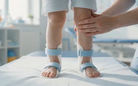
In spina bifida, foot, hip, knee and spine problems depend largely on the neurological level. We explain why regular orthopaedic follow-up matters, how treatment is planned and which warning signs to watch for.
On this page 8 sections
Spina bifida is a congenital condition in which the neural tube, the structure that forms the baby's spine and spinal cord, does not close completely during pregnancy. The most severe form, and the one that needs the most orthopaedic follow-up, is myelomeningocele, in which the spinal cord and its coverings protrude through the back in an open sac. Because leg strength and sensation depend on the level at which the spinal cord is affected, foot, hip, knee and spine problems also vary with that level. Regular spina bifida orthopaedics follow-up aims to protect mobility, limit the progression of deformities and pick up problems such as pressure sores and fractures early.
Spina bifida care is a team effort involving neurosurgery, urology, paediatrics, rehabilitation medicine and orthopaedics. This article explains why orthopaedic follow-up matters, the common problems and the warning signs families should know. Every child is different, and treatment is planned individually after examination and imaging.
What is spina bifida and what are its types?
The neural tube develops into the brain and spinal cord in the first weeks of pregnancy. When its lower part does not close fully, spina bifida results. There are three main types:
- Spina bifida occulta: a small gap at the back of one or more vertebrae, without the spinal cord or its coverings protruding. It usually causes no symptoms and is often found by chance on an X-ray taken for another reason.
- Meningocele: the coverings of the spinal cord protrude in a sac, but the cord itself usually stays in place, and neurological problems may be milder.
- Myelomeningocele: the spinal cord and nerves are within the sac. It needs closing after birth and is typically accompanied by leg weakness and reduced sensation, bladder and bowel problems and, often, a build-up of fluid in the brain (hydrocephalus).
Adequate folic acid intake before conception and in early pregnancy is known to reduce the risk of neural tube defects; it is worth discussing this with your obstetrician.
Why does the neurological level matter so much?
From an orthopaedic point of view, the most important piece of information is the child's neurological level: the level of the spinal cord down to which the muscles work. Muscles below that level are weak or paralysed and the skin has reduced feeling. The imbalance between working and non-working muscles gradually pulls joints in particular directions and causes deformity. The table below gives a general picture; individual children vary.
| Neurological level | Common orthopaedic problems | General outlook for mobility |
|---|---|---|
| Thoracic and high lumbar (L1–L2) | Hip and knee contractures, scoliosis and kyphosis | Mostly wheelchair; standing frames may be used in childhood |
| Mid lumbar (L3) | Hip subluxation or dislocation, knee problems, foot deformities | Limited walking in long orthoses; wheelchair for distance |
| Low lumbar (L4–L5) | Calcaneus (heel-walking) foot, valgus knee and ankle, outward shin twist | Walking with ankle-foot orthoses and, if needed, crutches |
| Sacral | High arches, claw toes, foot sores | Mostly independent walking |
The neurological level is monitored throughout childhood, because any new loss of strength can be the first sign of a treatable problem such as a tethered spinal cord, described below.
Foot problems and skin care in spina bifida
Most children with spina bifida develop a foot deformity at some point. Stiff clubfoot, vertical talus (a rocker-bottom foot), a calcaneus foot in which the child walks on the heel, and high-arched cavus feet are the main examples. The aim is a flat, plantigrade foot that fits into shoes and orthoses and stays as flexible as possible.
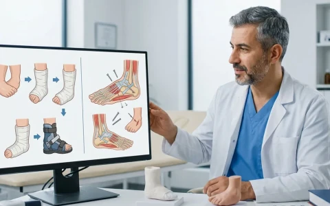
Ponseti casting can be used for clubfoot in these children too, although casts are applied with particular care because of reduced skin sensation, and relapse is more common. Our clubfoot treatment guide explains the method. When surgery is needed, procedures that preserve flexibility, such as tendon releases or transfers, are usually preferred, because a stiff foot without normal sensation is prone to pressure sores.
Reduced skin sensation is one of the most important issues in spina bifida. Parents are advised to check their child's feet every day for redness, blisters or sores, to watch the fit of shoes and orthoses closely, and always to test bath water by hand.
Hip and knee problems
Hip
Hip subluxation and dislocation are common, particularly at the mid-lumbar level. The muscles that bend the hip and pull it inwards work, while those that move it outwards and backwards are weak, so the femoral head is pushed out of the socket. Current practice focuses less on putting the hip back in the socket and more on preventing contractures and keeping the pelvis level. In many children, surgical reduction of a dislocated hip does not clearly change walking and carries a risk of stiffness or re-dislocation. Decisions are therefore individual, taking into account the neurological level, walking ability and whether one or both hips are affected.
Knee
A knee that cannot straighten fully (a flexion contracture) makes standing harder and can cause a crouched gait. In children with low lumbar levels who walk, knock-knee alignment and an outward twist of the shin bone can strain the knee and cause pain over the years. Orthoses help reduce this load, and for marked twisting a derotation osteotomy of the shin bone may be considered. For a broader view of childhood leg deformities, see our paediatric orthopaedics page.
Spinal curvature, fractures and conditions not to miss
Scoliosis and kyphosis: common especially at higher levels, they can disturb sitting balance, and progressive curves may need surgery. For background, see our article on scoliosis in children.
Fractures: reduced activity and sensation make bones thinner. Fractures can happen with minor force, for example during stretching or lifting, and particularly after a cast has been removed. The child may feel no pain; instead the leg becomes red, warm and swollen, sometimes with a fever. Because this can be mistaken for infection, an X-ray is important.
Tethered spinal cord: the cord becomes stuck to surrounding tissue and is stretched as the child grows. Loss of strength, worsening walking, a rapidly changing foot shape, accelerating scoliosis, back or leg pain and changes in bladder habits all raise suspicion and need neurosurgical assessment.
Latex allergy: it is more common in children with spina bifida, so the team should always be told in advance to ensure a latex-free environment for examinations and operations.
How are follow-up and treatment planned?
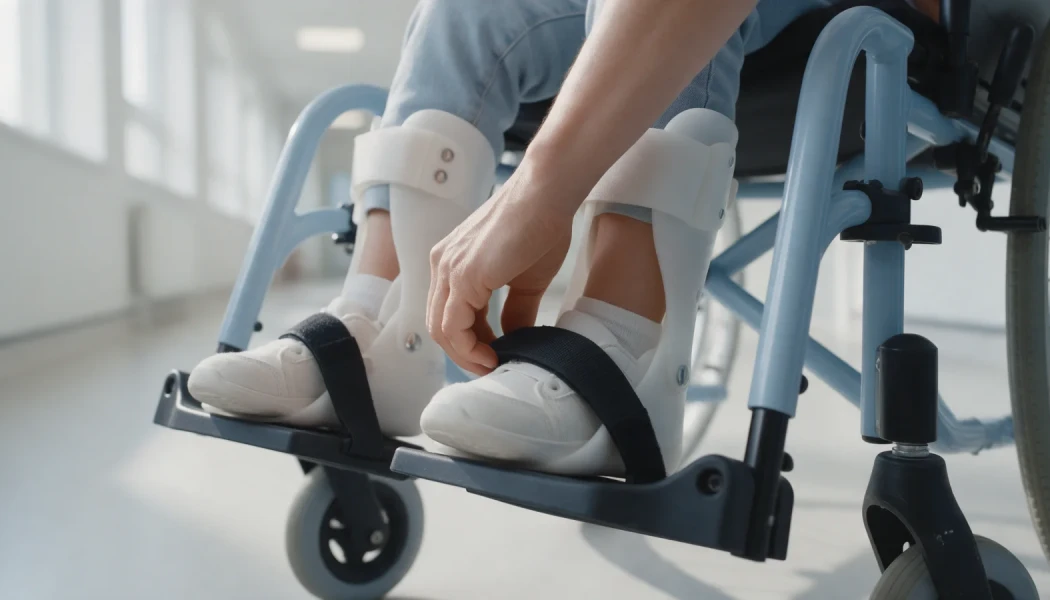
Check-ups are more frequent in infancy and usually every 6 to 12 months thereafter. Each visit reviews muscle strength, joint range of movement, the feet and skin, the hips and the spine, with X-rays when needed. Depending on the child's level, the programme may include physiotherapy, standing frames, ankle-foot or knee-ankle-foot orthoses, longer orthoses and a wheelchair.
Some principles guide surgical planning: keep the number of operations as low as possible, combine procedures where feasible, preserve joint flexibility and link every procedure to a functional goal. For children who walk, plantigrade feet that fit into orthoses are a priority; for wheelchair users, balanced sitting comes first. Extra care is taken during cast periods because of the fracture risk. For more on congenital and uncommon bone conditions, see our page on rare childhood limb deformities.
When should you see a doctor?
Alongside routine check-ups, seek prompt assessment if you notice:
- Unexplained redness, warmth or swelling of a leg (possible fracture)
- Redness, blistering or a sore on the feet or seating area that does not settle
- A decline in walking, muscle strength or the ability to use orthoses
- A rapid change in foot shape or in the curve of the back
- Changes in bladder or bowel habits together with changes in the legs
- Headache, vomiting, unusual drowsiness or irritability: these may indicate a shunt problem and need urgent attention
Orthopaedic care should be planned in close contact with the child's neurosurgeon, urologist and physiotherapist. Prof. Dr. Halil İbrahim Balcı is a faculty member at the Department of Orthopaedics and Traumatology of Istanbul Faculty of Medicine, working in paediatric orthopaedics and childhood deformities.
Key points
- The type of orthopaedic problem in spina bifida depends largely on the neurological level.
- For the feet, the goal is a flexible, plantigrade foot that fits into orthoses and shoes; skin should be checked daily.
- With hip dislocation, preventing contractures and keeping the pelvis level is often the priority; surgery is decided individually.
- Fractures can be silent; a red, warm, swollen leg always needs assessment.
- New weakness or rapidly worsening deformity should prompt investigation for a tethered cord.
Frequently Asked Questions
Spina bifida occulta usually causes no symptoms and needs no treatment; it is generally discovered by chance on an X-ray taken for another reason. However, if there are skin signs over the lower back, such as a tuft of hair, a dimple, a birthmark or a fatty lump, or symptoms such as leg weakness, a changing foot shape or bladder problems, further investigation of the spinal cord may be needed. Your doctor will advise on this.
This depends largely on the neurological level. Most children with sacral or low lumbar levels can walk, using orthoses and crutches if needed. At the mid-lumbar level, walking is usually possible with long orthoses over limited distances. At higher levels, a wheelchair is generally the main means of getting around. Muscle strength, joint contractures, weight and motivation also play an important part, so the outlook differs from child to child.
No, not always. Current practice prioritises preventing hip contractures and keeping the pelvis level. In many children, surgically reducing a dislocated hip does not clearly improve walking and carries a risk of stiffness or re-dislocation. Surgery may still be considered for some children, particularly those who walk and have a dislocation on one side only. The decision is made individually after examination and imaging.
Reduced muscle activity and less weight-bearing make the bones of the legs thinner, and because sensation is reduced the child may not notice strain or pain. Fractures can happen with minor force, especially after periods of immobility in a cast or after surgery. The usual sign is a red, warm and swollen leg rather than obvious pain. Standing programmes and careful handling can help protect bone health.
Latex allergy is more common in children with spina bifida than in the general population, probably because of frequent early exposure to latex-containing medical products. Reactions range from skin redness to severe allergic reactions. The healthcare team should therefore be told before every examination, procedure and operation so that latex-free materials are used. At home, avoiding latex balloons and toys can also help.
This article is for general information only and does not replace a medical examination, diagnosis or treatment. Treatment options and outcomes vary from person to person.
Related treatment area Rare Childhood Limb Deformities: Fibular Hemimelia, Femoral Deficiency