An osteochondroma is a benign, cartilage-capped bony lump that grows near a growth plate. We explain how a single lesion differs from the inherited multiple form (MHE) and when treatment is needed.

On this page 8 sections
- What is an osteochondroma and how does it form?
- Osteochondroma symptoms: what problems can it cause?
- What is multiple hereditary exostoses (MHE)?
- How is an osteochondroma diagnosed?
- Can an osteochondroma become cancerous?
- Osteochondroma treatment: monitoring or surgery?
- When should you see a doctor?
- Key points
An osteochondroma is a benign (non-cancerous) bony outgrowth with a cartilage cap that grows outwards from the surface of a bone, and it is the most common benign bone tumour found in children and teenagers. It usually appears as a hard lump near the knee or shoulder that often causes no pain at all. It is not cancer, and most osteochondromas simply need monitoring. When a child has many of these lumps rather than just one, the cause is usually an inherited condition called multiple hereditary exostoses (MHE).
Parents usually want to know whether the lump will keep growing, whether it needs an operation and whether siblings or future children could be affected. This guide answers those questions and explains how treatment decisions are made.
What is an osteochondroma and how does it form?
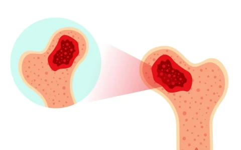
Near the ends of the long bones sit the growth plates (physes), the cartilage layers that make a child's bones longer. An osteochondroma is thought to begin when a small island of growth-plate cartilage ends up on the side of the bone and keeps growing away from the nearby joint. Over time this cartilage turns into bone, leaving a bony projection with a thin cartilage cap on top. The lump's outer shell and inner marrow are continuous with the bone it grows from, which is the key feature that allows diagnosis on an X-ray.
Osteochondromas come in two shapes:
- Pedunculated: a lump on a narrow stalk, rather like a mushroom or a thorn.
- Sessile: a flatter lump attached to the bone over a broad base.
The most common sites are around the knee (the lower end of the thigh bone and the upper end of the shin bone) and the upper end of the arm bone at the shoulder. The hip, pelvis, shoulder blade, forearm and ankle can also be affected. An osteochondroma grows as the child grows and usually stops growing once the growth plates close at skeletal maturity.
Osteochondroma symptoms: what problems can it cause?
Many osteochondromas cause no symptoms and are found by chance on an X-ray or noticed as a hard lump at bath time. When symptoms occur, they usually come from the lump pressing on or rubbing against nearby structures:
- Pain or a catching sensation with movement as a muscle or tendon slides over the lump
- A fluid-filled sac (bursa) forming over the lump, making it seem to enlarge suddenly
- A fracture through the stalk after a knock, causing sudden pain
- Pressure on nerves or blood vessels behind the knee or near the top of the fibula, causing numbness, tingling or weakness when lifting the foot
- Restricted joint movement when a large lump sits close to a joint
- Concerns about appearance, or rubbing from shoes and clothing
What is multiple hereditary exostoses (MHE)?
Multiple hereditary exostoses, also known as hereditary multiple osteochondromas, is an inherited condition in which osteochondromas develop on many bones. In most families it is linked to changes in the EXT1 or EXT2 genes and is passed on in an autosomal dominant pattern, which means each child of an affected parent has a one-in-two chance of inheriting it. Some children have no family history because the genetic change has arisen for the first time in them. MHE affects roughly 1 in 50,000 people and is usually recognised within the first ten years of life.
In MHE the problem is not simply the number of lumps. Lesions around the growth plates can disturb how bones lengthen and how they are shaped, so MHE is as much a growth and deformity condition as a tumour condition. We describe the wider approach to conditions like this on our page about rare childhood limb deformities.
Deformities that can occur with MHE
- Forearm deformity: a short ulna, a bowed radius, a wrist that drifts towards the little-finger side and, in some children, dislocation of the radial head at the elbow
- Knock knees (genu valgum) and outward tilting of the ankle
- Leg length difference and mildly reduced height
- Changes in the angle of the hip and reduced coverage of the hip joint
You can read more about correcting forearm bowing and shortening on our page on upper limb lengthening and deformity correction.
How is an osteochondroma diagnosed?
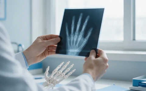
The first step, and often the only one needed, is a clinical examination and a plain X-ray, on which the lump merges seamlessly with the underlying bone. Further imaging is sometimes useful:
- MRI shows the thickness of the cartilage cap, its relationship to nearby muscles, nerves and blood vessels, and whether a bursa has formed.
- CT helps in areas that are difficult to assess on X-ray, such as the pelvis, shoulder blade or spine.
- Full-length standing leg X-rays are used in MHE to measure leg alignment and any difference in length.
A biopsy is not usually needed for an osteochondroma with a typical appearance. When MHE is suspected, the skeleton is assessed more widely, the family history is reviewed and genetic counselling is offered where appropriate.
Can an osteochondroma become cancerous?
This is the question parents ask most often. Malignant change in a single osteochondroma (to a secondary chondrosarcoma) is very rare. In MHE the likelihood is somewhat higher but remains low, and it usually occurs in adulthood. That is why regular review and body awareness matter, particularly for people with MHE. Signs that deserve attention include:
- A lump that keeps growing after the growth plates have closed
- New or increasing pain in adult life without an obvious cause
- A cartilage cap that appears clearly thickened on MRI in an adult
- Large lesions close to the trunk, for example in the pelvis, shoulder blade or hip
None of these signs means cancer on its own, but each one should be assessed by a team experienced in bone tumours. Our bone tumours page outlines how diagnosis and treatment are organised, and our article on bone tumours in children covers the symptoms worth watching for.
Osteochondroma treatment: monitoring or surgery?
An osteochondroma that causes no symptoms and looks typical usually needs nothing more than periodic review. Surgery is considered according to the problems the lump is causing. The table below summarises the general approach; for every child, the decision is made individually after examination and imaging.
| Situation | General approach |
|---|---|
| No symptoms, typical appearance | Observation and periodic review |
| Muscle or tendon irritation, recurrent pain, bursa | Surgical removal (excision) in suitable patients |
| Pressure on a nerve or blood vessel | Prompt assessment for surgical removal |
| Large lesion restricting joint movement | Excision, timed according to its distance from the growth plate |
| Angular deformity from MHE (knock knees, ankle tilt) | Guided growth while the child is still growing; corrective osteotomy if needed |
| Forearm deformity or leg length difference in MHE | Lesion removal, bone lengthening, epiphysiodesis or osteotomy |
| Growth or new pain in an adult | Further imaging and review by a bone tumour team |
Surgery aims to remove the whole lump with its cartilage cap and covering membrane, because tissue left behind can regrow. Lesions very close to a growth plate need careful planning to protect it, so in children with mild symptoms surgery is sometimes deferred until later in growth. Most children return to their daily routine fairly soon; the timing of a return to sport depends on the size and site of the bone removed.
Monitoring children with MHE as they grow
With MHE, follow-up means tracking bone alignment and limb lengths as the child grows, not just the lumps. Deformities can become more obvious during growth spurts, especially around the start of puberty, so review intervals depend on the child's age and findings. Typically:
- Height, limb lengths and joint movement are measured at each visit.
- The alignment of the forearm, knee and ankle is checked clinically and, when needed, with X-rays.
- While growth continues, the team considers whether a small procedure such as guided growth could stop a deformity from progressing.
- After puberty, the lesions to be followed in adult life are identified and the young person is told which changes to look out for.
In conditions such as MHE, where growth and deformity go hand in hand, it helps when paediatric orthopaedics, deformity surgery, radiology and, where needed, clinical genetics work together. Prof. Dr. Halil İbrahim Balcı developed his experience in deformity surgery and congenital bone anomalies at the Paley Institute in the USA, as a research fellow in 2014 and as an observer in 2019.
When should you see a doctor?
If you notice a hard lump that seems attached to your child's bone, it is sensible to have it checked by an orthopaedic surgeon even if it does not hurt. Do not put off an assessment if you notice:
- A lump that seems to have grown noticeably within a short time
- Pain that wakes your child at night or does not settle with rest
- Numbness, tingling or weakness in the leg or foot
- Difficulty fully bending or straightening a joint, or a limp
- Gradually increasing bowing of the forearm, knee or ankle
- A family history of MHE and more than one lump in your child
- A known osteochondroma that grows or starts to hurt in adult life
Key points
- An osteochondroma is a benign, cartilage-capped bony outgrowth that develops near a growth plate; it is not cancer.
- Most lesions that cause no symptoms are simply monitored; surgery is considered for problems such as pain, pressure on nerves or restricted movement.
- Multiple lumps suggest MHE, an inherited condition in which growth disturbances and deformities need close follow-up.
- Malignant change is rare, but growth or new pain in adulthood should always be assessed.
- Treatment is decided individually after examination and imaging, taking into account the child's age and remaining growth.
Frequently Asked Questions
Usually not. An osteochondroma tends to grow as the child grows and normally stops growing once the growth plates close. Shrinkage has occasionally been reported, but it is not something to count on. For lumps that cause no symptoms, the aim is to monitor their size, their relationship to the surrounding tissues and the child's growth at regular intervals. How often reviews are needed depends on the examination and X-ray findings.
When the lump is removed completely, together with its cartilage cap and covering membrane, regrowth at the same site is uncommon. The chance is higher if the lesion is removed at a young age while it sits right next to the growth plate, or if some tissue is left behind. In children with MHE, new lumps appearing on other bones are not a recurrence but part of the natural course of the condition, which is why follow-up continues.
MHE is inherited in an autosomal dominant pattern, so each child of an affected parent has a one-in-two chance of inheriting it. Severity can vary widely even within the same family: one person may have a few small lumps while another develops noticeable deformities. Genetic counselling can help families understand the risks, plan for the future and decide when children should be assessed.
Most children with an osteochondroma that causes no symptoms can take part in school sport and everyday activities. A stalked lump in an area that takes direct knocks can occasionally fracture or irritate surrounding tissues, so if pain, swelling or stiffness appears during contact sport, it is sensible to stop and have it checked. Which activities are suitable depends on where the lump is and how big it is, and should be agreed with your doctor.
There is no fixed age; the decision depends on the problems the lump is causing. Where there is pressure on a nerve or blood vessel, surgery may be brought forward whatever the child's age. For lumps with mild symptoms that sit close to a growth plate, surgery may be deferred until later in growth to protect the growth plate and reduce the chance of regrowth. The timing is planned individually after examination and imaging.
This article is for general information only and does not replace a medical examination, diagnosis or treatment. Treatment options and outcomes vary from person to person.
Related treatment area Rare Childhood Limb Deformities: Fibular Hemimelia, Femoral Deficiency