Most bone pain is not caused by a tumour, but certain features of pain and swelling should be assessed. We explain the warning signs, the diagnostic steps and when to see a doctor.

On this page 8 sections
- What is a bone tumour? Benign versus malignant
- Bone tumour symptoms: what to look out for
- Which pain and swelling should be taken seriously?
- How is a bone tumour diagnosed?
- Incidental bone lesions: is every finding a tumour?
- How is treatment planned after diagnosis?
- When should you see a doctor?
- Key points
The most common bone tumour symptoms are pain in a particular bone that lasts for weeks and gradually gets worse, pain that is noticeable at rest or at night, a hard swelling attached to the bone, and a fracture after only a minor knock. Most of these symptoms have causes other than tumours: the great majority of bone pain is due to strain, sports injuries or, in children, growing pains. What matters is recognising symptoms that do not settle within the expected time or that change in character, and having them examined and imaged without delay.
Most bone tumours, particularly in children and young people, are benign. Malignant tumours that start in bone (bone sarcomas) are rare compared with cancers overall. This article explains, without causing undue alarm, which signs deserve assessment and how the diagnostic process works.
What is a bone tumour? Benign versus malignant
A bone tumour is a general term for a lesion formed by cells growing in an uncontrolled way within bone tissue. Broadly, there are three groups:
- Benign tumours and tumour-like lesions: these include osteochondroma, enchondroma, osteoid osteoma, simple bone cyst, aneurysmal bone cyst and non-ossifying fibroma. They do not spread; some only need monitoring, while others need surgery. A few, such as giant cell tumour, are benign but can damage surrounding tissue, so they are treated more actively.
- Malignant tumours that start in bone: the most familiar are osteosarcoma and Ewing sarcoma, which are more common in teenagers and young adults, and chondrosarcoma, which mainly affects adults.
- Tumours that have spread to bone (metastases) and blood cell disorders: in adults over 40–50, a destructive lesion in bone is more often due to a cancer that has spread from another organ, or to multiple myeloma, than to a primary bone tumour.
Age, the bone involved and the part of the bone affected help the doctor rank the possibilities, but symptoms alone can never make the distinction.
Bone tumour symptoms: what to look out for
Pain
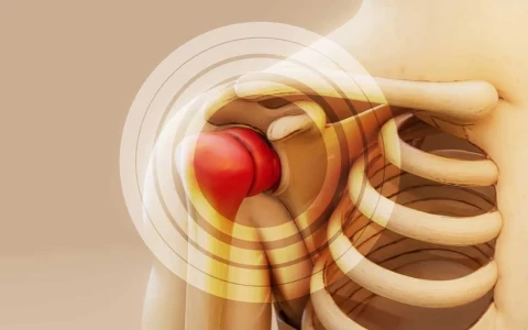
Pain is the most common symptom. At first it may come and go and be put down to overuse. Pain caused by a tumour is usually deep, aching and felt at one particular spot; over weeks it becomes constant, does not ease with rest and may be worse at night. Some benign tumours cause no pain at all.
Swelling or a lump
You may notice a hard swelling that seems fixed to the bone and does not move when pushed. Swellings that appear to be growing, particularly around the knee and shoulder, should be checked. Small, soft lumps that slide under the skin are usually not related to bone.
A fracture after a minor knock
When a tumour or cyst weakens the bone, it can break with a force that would not normally cause a fracture. This is called a pathological fracture and is sometimes the first sign of an underlying lesion.
Limping and reduced movement
Lesions near a joint can cause a limp, difficulty fully bending or straightening the joint, and wasting of the surrounding muscles.
General symptoms
Fever, unexplained weight loss, night sweats and tiredness are absent in most bone tumours, but they can occur with some tumours and with bone infections. When they accompany bone pain, assessment should be speeded up.
Which pain and swelling should be taken seriously?
The following signs do not mean cancer, but they do warrant an assessment by an orthopaedic surgeon and, in most cases, at least an X-ray:
- Bone pain lasting more than a few weeks that cannot be explained by a clear injury
- Pain that continues at rest or wakes you at night
- Pain fixed at one spot that increases in intensity over weeks
- Pain thought to be a sports injury that has not healed in the expected time, especially around the knee in teenagers
- A hard swelling attached to a bone that is getting bigger
- A fracture caused by a minor knock or an everyday movement
- Fever, weight loss or marked tiredness alongside bone pain
- New bone pain in someone who has previously been treated for cancer
Other common causes of bone pain
Pain around a bone has many far more common causes: overuse and strain, stress fractures, growing pains in children and growth-related pain at the front of the knee or the heel (such as Osgood-Schlatter and Sever's disease), bone and joint infections, and in adults, osteoarthritis and fractures related to osteoporosis. The aim of an assessment is therefore not only to look for a tumour but to find the real cause of the pain.
Our article on bone tumours in children: symptoms and early diagnosis looks at how these signs present in younger patients.
How is a bone tumour diagnosed?
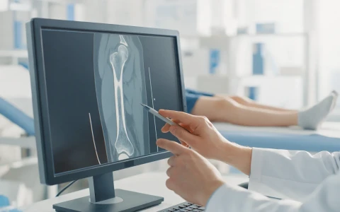
Diagnosis begins with a detailed history and physical examination. Imaging tests complement one another, and the diagnosis is confirmed, where necessary, by a biopsy: a tissue sample taken from the lesion and examined under the microscope.
| Test | What it shows |
|---|---|
| Plain X-ray | The first step; gives key information about the location and borders of the lesion and its effect on the bone |
| MRI | The extent of the lesion within the bone and surrounding soft tissue, and its relationship to nerves and blood vessels |
| Computed tomography (CT) | Details of the bone shell, calcification and fracture risk; also used to scan the lungs |
| Bone scan or PET-CT | Whether other bones or organs are involved, when this needs to be checked |
| Blood tests | Cannot make the diagnosis alone, but help with infection, blood disorders and general health |
| Biopsy | Confirms the type of lesion and whether it is benign or malignant |
When a malignant tumour is suspected, the biopsy should ideally be planned by the experienced team that will carry out definitive treatment, because the route taken for the biopsy can directly affect how any later operation is planned. Decisions about diagnosis and treatment are often made in multidisciplinary tumour boards that bring together orthopaedic surgeons, oncologists, radiologists, pathologists and radiation oncologists. You can read about this process on our bone tumours page.
Incidental bone lesions: is every finding a tumour?
A lesion seen in a bone on an X-ray or MRI taken for another reason is a common finding and is usually not a cause for concern. Lesions such as non-ossifying fibroma in children, and enchondroma or a bone island in adults, are frequently found by chance. Many of these, when they look typical, need no treatment at all, and some simply need to be checked at intervals.
If you are told about such a finding, the first step is to have the images and report reviewed by an orthopaedic surgeon with an interest in bone conditions, who can decide from the imaging features whether the lesion can be monitored or needs further tests. Reading report phrases online in isolation can cause unnecessary worry.
How is treatment planned after diagnosis?
Treatment depends on the type of tumour, its location and size, and the patient's age. For benign lesions, options range from observation to scraping out the lesion and filling the cavity with bone graft. For malignant tumours, surgery is planned together with chemotherapy and/or radiotherapy according to the tumour type.
Today, in suitable patients, the aim is to remove the tumour with safe margins while preserving the arm or leg (limb-sparing surgery). The missing bone can be reconstructed with tumour prostheses, bone grafts or bone transport using external fixators. We describe these methods in our article on surgical resection and reconstruction in bone tumours. Limb reconstruction with external fixators is also the subject of the Springer textbook based on Ilizarov principles that Prof. Dr. Halil İbrahim Balcı co-edited.
When should you see a doctor?
If any of the warning signs above has not improved within a few weeks, it is sensible to see an orthopaedic surgeon. A quicker assessment is needed if:
- An arm or leg suddenly becomes unusable after a minor strain, or you cannot put weight on it
- Back pain is accompanied by leg weakness, numbness or problems with bladder or bowel control (this needs emergency assessment)
- Pain over a bone comes with redness, warmth and fever
- A swelling grows noticeably within a short time
When you attend, it helps to note when the pain started, whether it wakes you at night and how it responds to painkillers, and to bring any previous scans with you.
Key points
- Most bone pain is not caused by a tumour, and most bone tumours are benign.
- Pain that lasts for weeks, gets steadily worse or is noticeable at rest or at night, and a growing swelling fixed to bone, should be assessed.
- An X-ray is usually the first step; MRI, CT and other tests complete the picture when needed.
- A definite diagnosis is made, when necessary, by a biopsy planned by an experienced team.
- Treatment is planned individually after examination and imaging, according to the tumour type and the patient's situation.
Frequently Asked Questions
Bone pain caused by a tumour is often described as a deep, aching pain felt at one particular spot. It may come and go at first, then become constant over weeks, fail to ease with rest and be worse at night. Its intensity and character vary with the type of tumour, however, and some benign tumours cause no pain at all. The features of the pain are therefore only a clue; the diagnosis is made by examination and imaging.
No single blood test can show a bone tumour. Tests such as a full blood count, inflammatory markers, alkaline phosphatase, LDH and, in adults, protein electrophoresis can point towards infection, blood disorders or certain tumour types. Normal results do not rule out a tumour, however, and abnormal results do not prove one. The key diagnostic steps are imaging and, when needed, a biopsy.
An X-ray is the first and an important step for most bone lesions, but very small or early lesions, those confined to the bone marrow, and those in areas that are hard to assess, such as the spine and pelvis, can be missed. If pain persists for weeks, gets worse or wakes you at night despite a normal X-ray, your doctor may request further imaging such as MRI. Persistent symptoms are a more important guide than a single normal X-ray.
The type of bone tumour varies markedly with age. Benign lesions are common in children and teenagers, while among malignant tumours osteosarcoma and Ewing sarcoma are seen mainly in adolescence and young adulthood. Chondrosarcoma mostly affects adults. In people over 40–50, a destructive lesion found in a bone is usually due to a cancer that has spread from another organ, or to multiple myeloma.
This is a worry that families often raise. A biopsy correctly planned by an experienced team is regarded as a necessary and safe way to confirm the diagnosis. The site and route of the biopsy are chosen so that the track can be removed during any later operation. That is why, when a malignant tumour is suspected, the biopsy is recommended at the centre that will plan definitive treatment.
This article is for general information only and does not replace a medical examination, diagnosis or treatment. Treatment options and outcomes vary from person to person.
Related treatment area Bone Tumours: Benign and Malignant Types, Diagnosis and Treatment