Osteoid osteoma is a small, benign bone tumour that can cause night-time bone pain in young people. Learn the typical symptoms, why CT is key to diagnosis and how it is treated today.
On this page 8 sections
Osteoid osteoma is a small, benign (non-cancerous) bone tumour that mostly affects children, teenagers and young adults. Its most typical symptom is pain felt at one particular spot in a bone that gets worse at night and can wake the person from sleep, and this pain often eases markedly with anti-inflammatory painkillers. Not every bone pain that worsens at night is an osteoid osteoma, but pain that lasts more than a few weeks, stays in one place and is worse at night should always be checked.
Osteoid osteoma does not spread and is not expected to turn malignant. Left untreated, however, it can cause months or even years of pain, a limp, muscle wasting and, in children, sometimes problems with growth. Today many patients can be treated with a minimally invasive procedure guided by computed tomography rather than open surgery.
What is an osteoid osteoma?
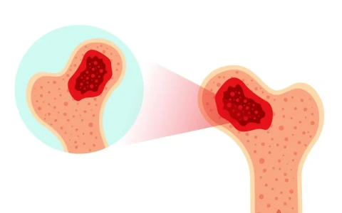
An osteoid osteoma is a small lesion that arises from bone-forming cells. At its centre is a focus called the nidus (Latin for nest), usually smaller than 1.5–2 cm, surrounded by bone that has become thickened and dense in reaction to it. The nidus produces large amounts of prostaglandins, substances involved in pain and inflammation. This explains the intensity of the pain, the widespread swelling in the surrounding tissue and the typical response to painkillers.
The tumour is most common between the ages of 5 and 25 and occurs more often in males. The bones most often affected are the thigh bone (femur), particularly the part near the hip, and the shin bone (tibia). The back of the spine and the bones of the hands and feet can also be involved, and less often the lesion sits inside a joint such as the hip. A related lesion, osteoblastoma, is larger (usually more than 2 cm), is more common in the spine and tends to cause less night pain.
Osteoid osteoma symptoms: why does night pain matter?
The pain may be mild and intermittent at first, then becomes constant over a few weeks and is often worst at night. Many patients and parents notice that it improves strikingly with anti-inflammatory painkillers (non-steroidal anti-inflammatory drugs). This response is an important diagnostic clue, but these medicines should not be taken for long periods without medical supervision, and masking the pain can delay diagnosis. Depending on where the lesion is, other symptoms may appear:
- In the long bones: a limp, muscle wasting in the affected leg and tenderness when pressing on one spot
- Inside a joint (especially the hip): fluid in the joint, stiffness and reduced movement, which can be mistaken for arthritis or inflammation of the joint lining
- In the spine: back pain, stiffness and a pain-related spinal curve (painful scoliosis)
- In the hands and feet: swelling, thickening and tenderness of a finger or toe
- Near a growth plate: a difference in leg length or angulation caused by increased blood flow
Growing pains or osteoid osteoma?
In many children who have pain at night, the cause is harmless growing pains. The table below is intended to give parents a general idea; the real distinction is made by examination and, where needed, imaging.
| Feature | Growing pains | Osteoid osteoma |
|---|---|---|
| Location | Usually both legs, in the muscles of the calf, thigh or back of the knee | Usually one side, at a specific spot in a bone |
| Timing | Evening or night; completely gone by morning | Worse at night; over time also present during the day |
| During the day | Child runs and plays normally, no limp | Limp or avoiding activities |
| Examination | Normal | Localised tenderness, possible muscle wasting |
| Pattern | Comes and goes, does not progress | Increases over weeks and becomes constant |
| Relief | Often settles with massage and warmth | Eases markedly with anti-inflammatory painkillers |
How is osteoid osteoma diagnosed?
Diagnosis starts with a detailed history of the pain and a physical examination, followed by imaging tests that are interpreted together:
- X-ray: may show thickening of the bone's outer layer with a small dark area inside it. Small or intra-articular lesions can be missed.
- Computed tomography (CT): thin-slice CT is the key test for showing the nidus and is also used to plan treatment.
- MRI: shows the extensive bone marrow oedema and soft tissue reaction around the lesion. When the nidus is small, this reaction can obscure it and make the picture look like an infection or another tumour, which is why MRI findings are usually read alongside CT.
- Bone scan: helps to locate the lesion in some cases.
Why can the diagnosis be delayed?
Because osteoid osteoma is so small, the first X-ray may look normal. In a sporty teenager the pain is often put down to overuse, and in a younger child to growing pains. Lesions inside a joint can resemble arthritis. The fact that painkillers keep the symptoms under control for long periods can also delay a visit to the doctor. For these reasons, the character and duration of the pain are often more telling than a single X-ray.
Conditions to rule out include a stress fracture, bone infection (such as a Brodie abscess), osteoblastoma and, less commonly, other tumours. Patients with typical findings may not need a separate biopsy, and a tissue sample can be taken during treatment if necessary. Our bone tumours page explains in more detail how benign and malignant bone lesions are assessed.
Osteoid osteoma treatment: what are the options?
Treatment is chosen according to the location and size of the lesion, the patient's age and their symptoms. The main options are:
- Monitoring with medication: some lesions are known to burn out on their own over several years. This can take a long time, however, and long-term use of painkillers carries a risk of side effects affecting organs such as the stomach and kidneys. It is therefore considered only in selected patients and under medical supervision.
- Radiofrequency ablation (RFA): under CT guidance, a thin needle is passed through the skin and the nidus is destroyed with controlled heat. The procedure is usually done under anaesthesia through a tiny entry point, and patients often go home the same day or after one night. For most accessible lesions it is now the usual first-line treatment.
- Other ablation techniques: microwave, laser and freezing (cryoablation) are also used, as is MRI-guided focused ultrasound in some centres.
- Surgical removal: may be preferred when the lesion lies very close to a nerve, blood vessel or the skin (for example in parts of the spine or in the fingers), when ablation is not technically suitable, when tissue is needed to confirm the diagnosis, or when pain returns after ablation. The aim is to remove the whole nidus, with bone graft or fixation added if needed.
We discuss why a timely diagnosis matters in our article on early diagnosis of bone tumours.
Recovery and follow-up after treatment
After radiofrequency ablation, pain usually starts to ease within a few days, although the treated area may be tender for a short time. Most people can return to school or work fairly soon. For lesions in weight-bearing areas such as the upper femur near the hip, strenuous sport and heavy activity may be restricted for a while to protect the bone; how long depends on the individual.
At follow-up, the team checks whether the pain has gone completely. If pain returns within months, part of the nidus may have survived, and repeat imaging is needed; the procedure can then be repeated or surgery considered. In children who developed a pain-related spinal curve or a leg length difference, these findings are monitored separately after treatment, and long-standing changes may need further treatment.
When should you see a doctor?
It is sensible to see an orthopaedic surgeon if you or your child have any of the following:
- Bone pain that wakes you from sleep and has lasted several weeks
- Pain fixed at one spot that is gradually increasing and becoming constant
- Pain that settles only with painkillers and returns when they are stopped
- A limp or thinning of the leg without any obvious injury
- A spinal curve or stiff back that has appeared together with pain
- Pain with swelling or redness over a bone, or with fever (this needs a quicker assessment to rule out infection)
Our article on bone tumours in children and early diagnosis describes other features of bone pain that deserve attention. Close cooperation between the orthopaedic surgeon, radiologists and interventional radiologists makes diagnosis and treatment planning more straightforward.
Key points
- Osteoid osteoma is a small, benign bone tumour of young people; it does not spread and is not expected to become malignant.
- The typical symptom is pain at one spot that worsens at night and eases markedly with anti-inflammatory painkillers.
- Thin-slice CT is the key diagnostic test; MRI findings alone can be misleading.
- In suitable patients, CT-guided radiofrequency ablation offers treatment without open surgery.
- Night pain lasting weeks, a limp or a painful spinal curve should be assessed with examination and imaging.
Frequently Asked Questions
No. Osteoid osteoma is a benign bone tumour: it does not spread to other parts of the body and is not expected to turn into a malignant tumour. Night pain and bone changes can, however, occur in other conditions too, so the diagnosis needs to be confirmed by examination and appropriate imaging. Once it is confirmed, treatment is planned according to how severe the pain is and where the lesion lies.
Some lesions have been reported to burn out on their own over several years. That process can take a long time, though, and in the meantime pain, poor sleep, a limp and muscle wasting can affect quality of life. Long-term use of painkillers also carries a risk of side effects. Whether to wait or to opt for ablation or surgery is therefore decided together with your doctor, taking into account where the lesion is and the patient's circumstances.
Because radiofrequency ablation is done through a tiny entry point, most patients get back to daily life quickly. Returning to school or desk-based work is often possible within a few days. Getting back to sport and heavy physical activity depends on whether the lesion is in a weight-bearing bone; with lesions in the upper femur near the hip, for example, a longer period of caution may be advised.
When the nidus is fully destroyed, pain returning is uncommon. If part of the lesion survives, the pain usually comes back within the following months with the same character as before. In that case the area is reassessed with CT, and the procedure can be repeated or surgical removal considered. That is why attending follow-up appointments and reporting any change in the pain to your doctor matters.
MRI shows the widespread oedema and soft tissue reaction around the lesion very clearly. The nidus itself is often smaller than 1–2 cm, so it can be lost among these changes, and the images may be interpreted as an infection, a stress fracture or another tumour. When osteoid osteoma is suspected, a thin-slice CT scan is therefore recommended, ideally reviewed by a team experienced in musculoskeletal radiology.
This article is for general information only and does not replace a medical examination, diagnosis or treatment. Treatment options and outcomes vary from person to person.
Related treatment area Bone Tumours: Benign and Malignant Types, Diagnosis and Treatment