Foot drop means the front of the foot cannot be lifted while walking, and over time it can lead to a fixed equinus deformity. We explain the causes, tests and treatment options, from braces to surgery.
On this page 8 sections
Foot drop is weakness or paralysis of the muscles that lift the ankle and toes, so the front of the foot cannot be raised properly while walking. Its most common causes are injury to the peroneal nerve, which runs around the outside of the knee, a nerve root compressed by a slipped disc in the lower back, and certain neurological conditions. Equinus deformity is a different but related problem in which the ankle becomes stiff in a pointed-down position; long-standing foot drop that is not managed can turn into equinus. Foot drop treatment is planned after examination, nerve tests and imaging, according to the cause, the likelihood of nerve recovery and whether any deformity is flexible or rigid.
This article will help you tell the two conditions apart and outlines the orthopaedic treatment options. For an overview of leg and ankle deformities in adults, see our adult leg deformity correction page.
What is foot drop and what causes it?
Lifting the foot is the job of the muscles at the front of the shin, chiefly tibialis anterior. A problem anywhere along the nerve pathway to these muscles can cause foot drop:
- Peroneal nerve injury: knee injuries and dislocations, fractures of the fibula, tight casts or braces, prolonged leg crossing or squatting, rapid weight loss and operations around the knee.
- Lower back causes: compression of the L5 nerve root by a disc prolapse or spinal stenosis.
- Sciatic nerve injury: hip injuries, hip surgery or injections into the buttock.
- Central nervous system conditions: stroke, multiple sclerosis, brain or spinal cord injury and cerebral palsy.
- Nerve and muscle conditions: diabetic nerve damage, inherited neuropathies such as Charcot-Marie-Tooth disease and some muscle disorders.
- After compartment syndrome: lasting damage to the muscles at the front of the leg.
Typical signs include the foot slapping on the ground, a "high-stepping" gait in which the knee is lifted higher than usual so the toes clear the floor, tripping on stairs and kerbs, and frequent ankle sprains. Peroneal nerve injury may also cause numbness over the top of the foot.
What is equinus deformity and how does it differ from foot drop?
In equinus deformity the calf muscles or Achilles tendon have shortened, so the ankle cannot be lifted far enough; in more severe cases it cannot even reach a right angle to the leg. The heel does not reach the ground fully, weight shifts onto the front of the foot and the knee may be pushed backwards when walking. Long-standing foot drop is only one of several common causes:
| Feature | Foot drop | Equinus deformity |
|---|---|---|
| Main problem | Weakness of the muscles that lift the foot | Shortening and stiffness of the calf and Achilles tendon |
| Can the foot be lifted by hand? | Usually yes, at first | Only partly, or not at all |
| Common causes | Nerve injury, disc prolapse, neurological conditions | Long-standing foot drop, spasticity, residual clubfoot, fractures and compartment syndrome, long immobilisation, bone lengthening |
| Focus of treatment | Treat the cause, support the foot, restore function | Lengthen the shortened tissues, correct the ankle position |
In children, stiffness remaining after treatment of congenital clubfoot can also lead to equinus; this is covered in our clubfoot treatment guide.
How is foot drop diagnosed?
Examination assesses muscle strength, sensation, reflexes and walking. The Silfverskiöld test compares how far the ankle can be lifted with the knee bent and with it straight, which shows whether the tightness lies only in the gastrocnemius (the upper calf muscle) or in the whole Achilles unit. Other commonly used tests are:
- Electromyography (EMG) and nerve conduction studies: these show where the nerve is damaged and how severely; they are usually most informative a few weeks after symptoms begin.
- MRI: of the lower back if a spinal cause is suspected, or of the knee region if local compression is suspected.
- Ultrasound: used in some centres to image the nerve directly.
- Standing X-rays of the foot and ankle: to assess bone deformity and the condition of the joints.
Investigating the underlying cause is often shared with neurologists, spinal surgeons or rehabilitation specialists.
Non-surgical foot drop treatment
The first step is to remove the cause where possible, for example adjusting a cast or brace that is pressing on the nerve, treating a disc prolapse or controlling an underlying condition. Some peroneal nerve palsies caused by compression recover over weeks or months. Meanwhile, the aim is to protect the foot and prevent equinus from developing:
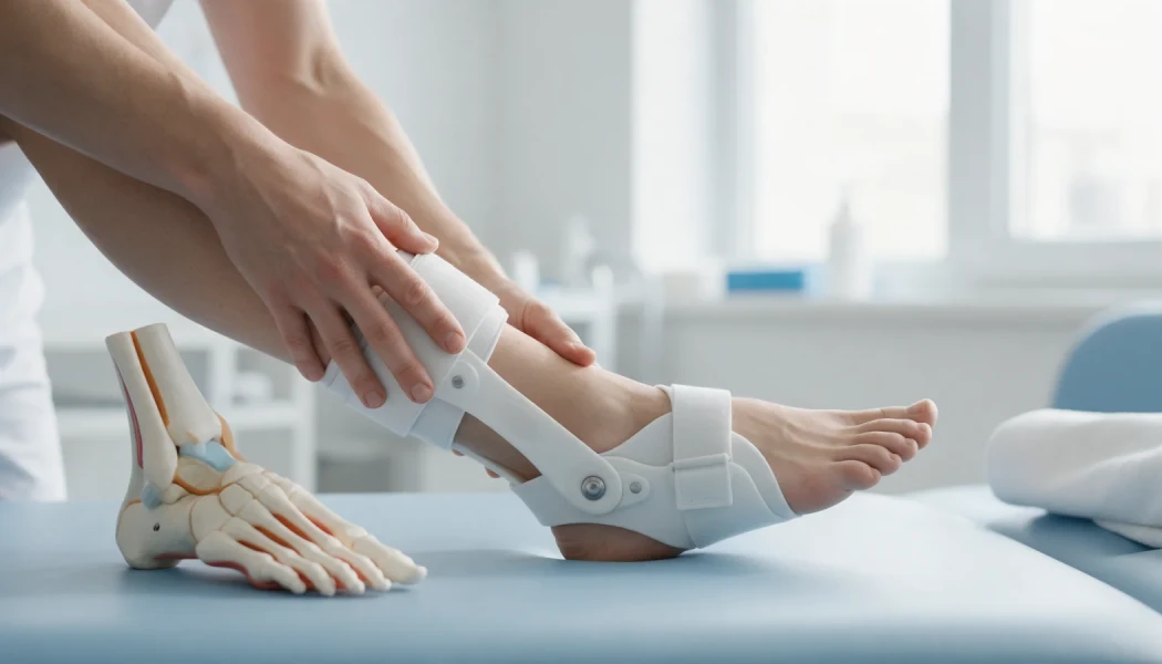
- Ankle-foot orthosis (AFO): holds the foot up while walking, reduces tripping and helps prevent the calf from shortening.
- Physiotherapy: daily calf stretches, strengthening, balance and gait training.
- Night splints: keep the ankle at a right angle during sleep to help prevent tightening.
- Serial casting: can gradually stretch the calf in mild to moderate, flexible equinus.
- Electrical stimulation and spasticity treatments: may be used in central causes such as stroke or cerebral palsy, on the advice of the relevant specialists.
When is surgery considered?
Nerve surgery
When a nerve is trapped or injured, release of the nerve, repair, nerve grafting or nerve transfer may be considered. Timing matters for these procedures, and they are usually planned within the first months, by teams experienced in peripheral nerve surgery.
Tendon transfer
When nerve recovery is not expected, the tendon of tibialis posterior, a muscle that normally turns the foot inwards, can be rerouted through the membrane between the two leg bones to the top of the foot. The muscle then takes over the task of lifting the foot. In suitable patients this can reduce the need for a brace, but it requires a strong donor muscle and a flexible ankle, and the muscle learns its new role over months of rehabilitation.
Correcting equinus deformity
Flexible or moderately stiff equinus may be treated by lengthening the covering of the calf muscle (gastrocnemius recession), lengthening the Achilles tendon and, if needed, releasing the back of the ankle capsule. The choice depends on the Silfverskiöld test and the examination findings.
Gradual correction with the Ilizarov method
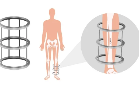
In severe, long-standing deformity, in scarred tissues or after previous surgery, correcting the foot in a single operation can place too much tension on the skin, blood vessels and nerves. In these situations a circular external fixator can bring the ankle gradually into the right position over several weeks, combined with bone correction if needed. The principles are explained on our Ilizarov and limb reconstruction surgery page, and our article on deformities that can be corrected with the Ilizarov technique gives further examples. Ilizarov techniques and deformity surgery are among the areas Prof. Dr. Halil İbrahim Balcı studied at the Paley Institute.
Joint fusion (arthrodesis)
Where there is advanced arthritis, marked instability, or other methods are not suitable, fusing the ankle or the hindfoot joints may be an option to reduce pain and give a stable, flat foot to stand on.
What does recovery involve?
After soft-tissue surgery the foot is usually protected in a cast or boot for a few weeks before moving on to an orthosis and physiotherapy. After a tendon transfer it can take months for the muscle to learn to lift the foot. With gradual correction in a frame, the frame typically stays on for weeks to months, and a brace may then be used to hold the corrected position. With every method, recovery time and the function achieved vary with the underlying cause and the individual.
When should you see a doctor?
- Foot drop has come on suddenly, especially with facial drooping, arm weakness or slurred speech (emergency)
- Back pain is accompanied by loss of bladder or bowel control or numbness around the groin and buttocks (emergency)
- After a knee injury, a fracture or a cast you cannot lift your foot, or the top of your foot feels numb
- Weakness is getting worse, or affects both feet
- You trip frequently, or your heel no longer reaches the ground
Key points
- Foot drop is caused by weakness of the muscles that lift the foot; equinus is stiffness of the ankle in a pointed-down position due to a tight calf and Achilles tendon.
- Peroneal nerve injury, disc prolapse and neurological conditions are common causes of foot drop.
- Orthoses, stretching and physiotherapy support walking and help prevent equinus from developing.
- Nerve surgery, tendon transfer, soft-tissue lengthening and gradual Ilizarov correction are surgical options considered in suitable patients.
- The right treatment is chosen after examination, nerve tests and imaging; results vary from person to person.
Frequently Asked Questions
It depends on the cause. Some peroneal nerve palsies due to compression, and some weakness caused by a disc prolapse, recover over weeks or months once the pressure is relieved. When the nerve has been completely divided or compressed for a long time, recovery may be limited. Progress is followed with regular examinations and, where needed, repeat EMG studies.
An orthosis is used while the nerve has a chance to recover, to reduce tripping and prevent the calf from tightening. As the nerve improves the need for the brace may lessen, while if recovery is not expected, long-term use or surgical options are discussed. The type of orthosis and how long it is worn are tailored to your muscle strength and walking pattern.
In suitable patients a tibialis posterior tendon transfer can reduce the need for a brace, and some people are able to walk without one. The outcome, however, depends on the strength of the transferred muscle, the flexibility of the ankle, the extent of any nerve damage and commitment to rehabilitation, and it varies from person to person. Realistic goals should be discussed with your surgeon before the operation.
Mild, flexible equinus can improve considerably with regular stretching, night splints and, where needed, serial casting. In long-standing, rigid deformity, or when scar tissue is involved, exercise alone is often not enough and surgical options are considered. The degree of deformity is assessed by examination and, where helpful, imaging, which guides the choice between these approaches.
New or worsening foot weakness due to a disc prolapse needs prompt assessment, because timing can matter when deciding on treatment. If there is also loss of bladder or bowel control, numbness around the groin and buttocks, or weakness in both legs, it is an emergency and you should go to an emergency department without delay.
This article is for general information only and does not replace a medical examination, diagnosis or treatment. Treatment options and outcomes vary from person to person.
Related treatment area Adult Leg Deformity Correction: Bow Legs and Knock Knees