The Masquelet technique repairs large gaps in bone over two operations. We explain how it works, who it may suit, what recovery involves and how it compares with Ilizarov bone transport.
On this page 8 sections
- What is a bone defect and why does it not heal on its own?
- How is the Masquelet technique performed?
- Who might the Masquelet technique be suitable for?
- Masquelet technique or bone transport: how do they compare?
- What are the possible risks and problems?
- What should patients expect during treatment?
- When should you contact your doctor?
- Key points
The Masquelet technique (the induced membrane technique) is a two-stage method for rebuilding large gaps in bone caused by trauma, infection or tumour surgery. In the first operation a temporary bone cement spacer is placed in the gap, and the body forms a biologically active membrane around it; in the second, the spacer is removed and the membrane is filled with bone graft. Whether the method is suitable depends on the location and length of the defect, the infection status, the soft tissues and the patient's general health.
The technique is named after the French surgeon Alain-Charles Masquelet and has been used widely around the world since the 2000s. Alongside bone transport with the Ilizarov method, it is now regarded as one of the main options for large bone defects.
What is a bone defect and why does it not heal on its own?
A bone defect is a gap left between two bone ends when a section of bone has been lost. It can result from bone fragments lost in a severe open fracture, removal of infected bone, excision of a tumour or previous non-union operations that did not succeed. Small gaps can close through the bone's natural healing capacity, but beyond a certain size the bone cannot bridge the gap by itself. Bone graft placed directly into such a gap may also be resorbed if its blood supply is poor. The Masquelet technique addresses exactly this problem by creating a biological environment in which the graft can survive.
The main methods used for large bone defects are the Masquelet technique, bone transport with the Ilizarov method, transfer of living bone such as a vascularised fibula graft and, in suitable cases, acute shortening followed by lengthening. Which path is chosen depends on the length and location of the defect, the infection status and the patient's own priorities.
How is the Masquelet technique performed?
Stage one: debridement, spacer and stabilisation
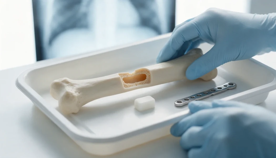
In the first operation, dead or infected bone and soft tissue are removed back to healthy tissue (debridement), and samples are taken for microbiology. The gap is filled with a spacer made of bone cement (PMMA), with antibiotics added where needed. The bone is stabilised with an external fixator, a plate or an intramedullary nail. If the soft-tissue cover is inadequate, a tissue transfer (flap) may be planned with plastic surgeons.
The interval: forming the induced membrane
The body treats the cement as a foreign body and, over a few weeks, builds a thin, well-vascularised membrane around it. This "induced membrane" releases growth factors that support bone healing and protects the graft that will be placed later, acting rather like a biological chamber. The interval between the two stages is classically around 4 to 8 weeks, and it may be longer while infection is still being treated. Before stage two, the wound and blood tests are reviewed to confirm that infection is under control.
Stage two: bone grafting
In the second operation the membrane is opened carefully, the spacer is removed and, with the membrane preserved, the space is packed with the patient's own cancellous (spongy) bone. This is usually harvested from the pelvis or, with a dedicated reaming system, from the inner canal of the femur; in large defects it may be extended with other graft materials. Our article on bone grafts used in non-union explains the options in more detail. The membrane is then closed and the fixation is reassessed.
Consolidation
Over the following months the graft gradually matures into solid bone. Progress is monitored with X-rays and, when needed, CT scans, and weight-bearing on the leg or loading of the arm is increased step by step as consolidation advances. How long full consolidation takes depends on the length of the defect, the bone involved, the fixation method and the individual's biology. Rehabilitation to rebuild muscle strength and joint movement continues for some time after the bone has become solid.
Who might the Masquelet technique be suitable for?
In suitable patients, the method may be considered for:
- loss of a section of bone after an open fracture
- gaps left after extensive debridement of an infected non-union (see complex and infected non-union)
- removal of an affected section of bone in chronic bone infection (osteomyelitis)
- defects left after a bone tumour has been removed
- selected patients in whom an earlier reconstruction has not worked
The main factors in the decision are the quality of the soft-tissue cover, whether infection can be controlled, the blood supply to the area, smoking, conditions such as diabetes, nutrition, and the patient's ability to commit to a long course of treatment. Where infection cannot be controlled, soft-tissue cover cannot be achieved or two operations are not appropriate, other options may be preferable.
Masquelet technique or bone transport: how do they compare?
The two methods most often compared for large bone defects are the Masquelet technique and bone transport using Ilizarov principles. In bone transport, a segment of healthy bone is cut and moved into the gap with a frame at about 1 mm a day, and new bone forms in the space it leaves behind.
| Feature | Masquelet technique | Bone transport (Ilizarov) |
|---|---|---|
| Principle | Create a membrane with cement, then fill the gap with graft | Move a bone segment gradually to generate new bone |
| Number of operations | At least two planned operations | Frame application and removal; extra surgery at the docking site may be needed |
| External fixator | Can be done with internal fixation; frame time is usually shorter | Frame stays on for months, in proportion to the length of the defect |
| Bone graft | A large volume of graft is needed | No large graft; a small graft may be added at the docking site |
| Length and alignment | Does not correct shortening or deformity on its own | Lengthening and deformity correction can be done at the same time |
| Typical problems | Graft resorption or incomplete union, pain at the donor site | Pin-site infection, joint stiffness, delayed healing at the docking site |
These methods are complementary rather than competing, and in some patients they are combined. Our article on external fixators in non-union covers the role of frames in more depth. Prof. Dr. Halil İbrahim Balcı is a co-editor of the Springer book on external fixator applications according to Ilizarov principles (2018).
What are the possible risks and problems?
As with any reconstruction, problems can arise with the Masquelet technique:
- recurrence of infection, needing further debridement
- partial resorption of the graft or incomplete union, needing re-grafting
- temporary pain or tenderness at the graft donor site
- fracture of the reconstructed segment or problems with the fixation hardware
- joint stiffness and muscle weakness after long periods of restricted activity
How far these risks apply varies from person to person, and they should be discussed in detail before surgery. For the general principles of treating bones that fail to heal, see our non-union surgery page.
What should patients expect during treatment?
Treatment with the Masquelet technique can take from several months to more than a year. During this time patients usually have two separate hospital stays, wound care, antibiotic treatment as planned by the team and regular follow-up appointments. Physiotherapy matters from the start, to keep the joints moving, maintain muscle strength and learn to walk safely within weight-bearing limits. Stopping smoking, keeping blood sugar under control and eating enough protein all support bone healing. While mobility is restricted, following the team's advice on preventing blood clots (deep vein thrombosis) is also important. Fatigue and worry are common during a long treatment, and family support and open communication with the team make the journey easier.
When should you contact your doctor?
During or after treatment, contact your team without waiting for the next planned appointment if you notice:
- fever, shivering or feeling generally unwell
- redness, increasing swelling, discharge or opening of the wound
- pain that is getting worse or is new
- loosening, discharge or an unpleasant smell around fixator pins
- a new bend, shortening or a sense of movement in the limb
- numbness, coldness or colour change in the foot or hand
Key points
- The Masquelet technique rebuilds large bone defects in two stages, filling a cement-induced biological membrane with bone graft.
- The two stages are usually a few weeks apart; infection control determines the timing.
- Compared with bone transport, frame time is usually shorter, but a large amount of graft is needed and shortening is not corrected on its own.
- Suitability depends on the defect, the soft tissues, infection status and general health.
- Consolidation takes months, and results vary from person to person.
Frequently Asked Questions
In the classic approach the two stages are around 4 to 8 weeks apart, which gives the membrane around the spacer time to mature. If infection treatment is still under way, or the soft tissues need longer to heal, the interval can be extended. The timing of the second operation is decided by reviewing the wound, blood tests and the patient's general health together.
The graft is usually taken from the patient's own pelvis or, using a dedicated reaming system, from the inner canal of the femur. The donor site may be sore, tender or bruised for a while after surgery, and these symptoms usually settle over the following weeks. Pain relief is planned by the treating team, and when a large volume is needed, other graft materials can be used to supplement it.
Most patients start moving with crutches soon after surgery, but how much weight the leg can take depends on the fixation method, the location of the defect and how far the graft has consolidated. Weight-bearing is usually increased gradually according to the X-rays. Moving to full weight-bearing often takes months, and the timescale varies from person to person.
In the Masquelet technique the cement is not a bone substitute but a temporary filler that stimulates the membrane to form. In the classic approach it is therefore removed at the second stage and replaced with bone graft. In the uncommon situations where a second operation is not possible, the options are considered separately with the team, taking the patient's health and wishes into account.
Yes, in selected children, for example after tumour surgery, bone infection or some congenital non-unions. Children generally have a greater capacity for bone healing, but protecting the growth plates and anticipating length differences that may appear with growth are additional considerations in planning. The decision is made by a team with experience in paediatric orthopaedics.
This article is for general information only and does not replace a medical examination, diagnosis or treatment. Treatment options and outcomes vary from person to person.
Related treatment area Revision Surgery for Complex and Infected Nonunion