Congenital pseudarthrosis of the tibia is a rare condition in which a weak segment of a child's shin bone fractures and does not heal. We explain the signs, the NF1 link and current treatment principles.
On this page 8 sections
- What is congenital pseudarthrosis and what happens in the bone?
- How is congenital pseudarthrosis of the tibia linked to NF1?
- How is congenital pseudarthrosis of the tibia diagnosed?
- What can be done before a fracture occurs?
- What principles guide surgery for congenital pseudarthrosis of the tibia?
- What happens in the long term after surgery?
- When should you see a doctor?
- Key points
Congenital pseudarthrosis of the tibia is a rare condition in which a weak, bowed segment of a child's shin bone (tibia) breaks and does not heal on its own, leaving a "false joint" in the bone. It is often first noticed in infancy as a forward and outward bow in the lower leg, and the fracture usually occurs in the first years of life, sometimes after only a minor stumble. Treatment aims to achieve union, prevent the bone from breaking again and protect the alignment and length of the leg.
Congenital pseudarthrosis of the tibia (CPT) is reported in roughly 1 in 140,000 to 1 in 250,000 births. The word pseudarthrosis is also used for fractures that fail to heal in adults, but the congenital form has different causes and needs a different approach. If you are reading about adult non-union, our article on non-union (pseudarthrosis) treatment options may be more relevant.
What is congenital pseudarthrosis and what happens in the bone?
In CPT a segment of the tibia, usually in its lower third, does not develop normally. The membrane covering the bone (periosteum) is thickened, and the bone is surrounded by abnormal fibrous tissue with little ability to form bone. This tissue has a limited blood supply and poor healing capacity. As a result, a fracture that would heal within weeks in another child does not unite, and over time the bone ends become thin and tapered. The fibula, the slender bone beside the tibia, is often involved as well.
How is congenital pseudarthrosis of the tibia linked to NF1?
Neurofibromatosis type 1 (NF1) is a genetic condition that can affect the skin, nerves and bones. Its most recognisable signs are light brown "café-au-lait" patches on the skin, freckling in the armpits or groin, and soft lumps arising from nerve sheaths (neurofibromas). The diagnosis rests on defined clinical criteria, supported by genetic testing where needed; six or more distinct café-au-lait patches, for example, is one of these criteria.
- A substantial proportion of children with CPT have NF1; published series report around half or more.
- However, only a small minority of children with NF1 develop tibial involvement, so a diagnosis of NF1 does not mean a child will have a bone problem.
- A child diagnosed with CPT is assessed for NF1 by paediatric neurology, genetics and eye specialists, and a child with NF1 and a bowed leg is followed by an orthopaedic surgeon.
- In some children CPT occurs with other bone conditions such as fibrous dysplasia, or with no associated condition at all.
How is congenital pseudarthrosis of the tibia diagnosed?
Is every bowed leg a cause for concern?
No. Most bowing seen in babies and toddlers is harmless. The direction of the bow, whether one or both legs are affected and the X-ray appearance help tell the types apart:
| Type of bowing | Features | Usual course |
|---|---|---|
| Physiological bow legs | Both legs, at knee level, most obvious when toddlers start walking | Usually straightens with growth |
| Posteromedial bowing | Present at birth, the foot often sits turned upwards | Tends to improve over the years; a length difference may remain |
| Anterolateral bowing | Usually one leg, lower tibia; may be associated with NF1 | Does not correct itself; carries a risk of fracture and pseudarthrosis, needs close follow-up |
X-ray findings and classification
Examination and X-rays are the foundation of diagnosis. At the apex of the bow the X-ray may show a narrowed marrow canal, dense (sclerotic) bone, cyst-like areas or a fracture with tapered ends. MRI is used in some children to show how far the abnormal tissue extends. Leg length and ankle alignment are monitored with standing full-length leg X-rays.
The Crawford classification, based on the X-ray appearance, is widely used:
- Type I: anterolateral bowing with thick cortical bone and a narrow but open marrow canal.
- Type II: bowing with a narrowed, sclerotic marrow canal.
- Type III: a cyst-like area at the apex of the bow; a pre-fracture stage.
- Type IV: an established fracture and false joint, often with thinned bone ends.
Other systems, such as the Paley classification, which takes into account involvement of the tibia and fibula and the gap between the bone ends, also help with surgical planning.
What can be done before a fracture occurs?
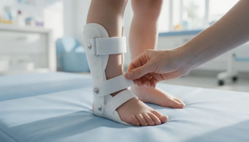
When anterolateral bowing is found but the bone has not yet broken, the aim is to protect the weak segment. Once the child begins to stand and walk, a custom-made brace that encloses the leg is usually recommended and worn throughout the day. Cutting the bone to straighten the bow (osteotomy) is generally avoided in this condition, because it can create a non-union. In selected children, operations to reinforce the bone before a fracture occurs may be discussed; the decision depends on the degree of bowing and the X-ray findings.
A few practical points help families living with a brace:
- Check the skin every day when the brace comes off, looking for redness, blisters or sore spots.
- Use a seamless cotton sock or liner under the brace.
- Have the fit reviewed every few months, as children grow quickly.
- Let nursery, school and other carers know why the brace matters and when it should be worn.
What principles guide surgery for congenital pseudarthrosis of the tibia?
Once a pseudarthrosis has developed, treatment is surgical. Rather than a single standard operation, it combines several principles:
- Removing the abnormal tissue: the thickened periosteum and surrounding fibrous tissue are excised back to healthy bone.
- Stable fixation: the bone ends are held, and compressed where needed, with a rod inside the bone (sometimes crossing the ankle), a circular external fixator (Ilizarov) or both together.
- Biological support: bone graft taken from the child's own pelvis and periosteal graft from a healthy area encourage healing. Some protocols also add medicines that influence bone metabolism, at the treating team's discretion.
- Bridging the tibia and fibula: combined techniques aimed at creating a solid bony bridge between the two bones, known as "cross-union", have been reported with encouraging results in recent years.
- Alternative methods: in selected children a vascularised fibula graft or bone transport with the Ilizarov method may be used.
After surgery the bone is protected for a period with a cast, a brace or the frame, depending on the method used. Progress towards union is followed with regular X-rays, and weight-bearing is increased step by step according to these findings. The timing of surgery depends on the child's age, the size of the bone and the state of the fracture. Our page on complex and infected non-union describes the general approach to difficult and recurrent non-unions. Results vary from child to child.
What happens in the long term after surgery?
Even after the bone has healed, the risk of refracture continues until the end of growth and sometimes beyond. For this reason the rod inside the bone is often left in place for a long time, and a protective brace may be worn for years. Follow-up checks leg length, any outward tilt of the ankle (valgus) and the position of the fibula, and later surgery to equalise length or correct alignment may be needed. In the rare situations where union cannot be achieved despite several operations, options that prioritise function with a prosthesis are discussed openly with the family. Throughout this long journey, keeping school and play as normal as possible matters a great deal.
When should you see a doctor?
- You notice a clear forward and outward bow in your baby's shin
- Your child has several café-au-lait patches on the skin together with a bowed leg
- After a minor fall your child refuses to put weight on the leg or starts to limp
- A tibial fracture has been diagnosed and it is not healing in the expected time
- The brace rubs, irritates the skin or is starting to feel too small
Prof. Dr. Halil İbrahim Balcı's work at the Paley Institute focused on congenital bone anomalies and Ilizarov techniques; conditions such as CPT are covered within rare childhood limb deformities.
Key points
- Congenital pseudarthrosis of the tibia is a fracture in a congenitally weak segment of the shin bone that does not heal on its own.
- Anterolateral bowing is the key early sign; most other bowing in babies is harmless.
- Many children with CPT have NF1, so the two conditions are assessed together.
- Bracing protects the bone before a fracture; afterwards, surgery combines excision, stable fixation and biological support.
- Because refracture remains possible, follow-up continues until the end of growth, with a plan tailored after examination and imaging.
Frequently Asked Questions
In some babies a forward and outward bow, or even a mobile fracture, is visible at birth. In many others there is only a mild bow at first, and the fracture develops in the early years, often after the child starts walking. An X-ray for any baby with a noticeable bow in the shin helps to identify the higher-risk type of bowing early, before a fracture has had the chance to occur.
Most children with NF1 never develop tibial involvement. Even so, the shape of the legs should be checked at routine NF1 reviews. If you notice a forward and outward bow of the shin, one leg looking shorter or a limp, it is worth arranging a paediatric orthopaedic assessment. Bowing that is identified early can be protected with a brace and monitored closely over time.
Before a fracture, the brace is usually worn during the day from the time the child starts standing, and it is renewed as they grow. After surgery, even once the bone has united, a protective brace is often recommended for years, sometimes until the end of growth, to reduce the risk of refracture. The duration is reviewed at follow-up according to the X-rays, the child's age and their activity level.
Once the bone has united and muscle strength has returned, many children can enjoy activities such as walking, swimming and cycling. Sports involving sudden twisting or collisions are considered separately because of the risk of refracture. Which activities are suitable is decided individually, based on the X-rays, the rod and brace, and the child's age and maturity.
This article is for general information only and does not replace a medical examination, diagnosis or treatment. Treatment options and outcomes vary from person to person.
Related treatment area Rare Childhood Limb Deformities: Fibular Hemimelia, Femoral Deficiency