Tarsal coalition is an abnormal connection between foot bones present from birth, often noticed in adolescence through recurrent sprains and pain. We explain the symptoms, diagnosis and treatment options.
On this page 8 sections
Tarsal coalition is an abnormal bridge of bone, cartilage or tough fibrous tissue joining two or more of the bones in the back and middle of the foot (the tarsal bones). It is present from birth but usually causes no symptoms in early childhood. As the bridge stiffens during adolescence, it restricts foot movement and can cause recurrent ankle sprains, foot pain that worsens with activity and a rigid flat foot. Diagnosis is made with examination and imaging; treatment starts with non-surgical measures, and surgery is considered if symptoms persist.
In teenagers brought to clinic because they "keep going over on their ankle", tarsal coalition is one of the causes that can be missed. This article explains what it is, the symptoms it causes, how it is diagnosed and the treatment options, in terms families and young people can follow.
What is tarsal coalition and why does it happen?
The bones of the foot develop in the womb by separating from a single mass of tissue. If this separation is incomplete between two bones, a bridge remains. Tarsal coalition can run in families, which suggests a genetic tendency. It is more common than once thought, but many people never have symptoms, and in about half of cases both feet are affected.
The bridge may be fibrous, cartilaginous or bony. Fibrous and cartilaginous bridges are more flexible in childhood but begin to turn to bone in adolescence, which is when the foot stiffens and symptoms tend to appear. The two most common types are:
- Calcaneonavicular coalition: a bridge between the heel bone (calcaneus) and the navicular bone in the midfoot. Symptoms often start between about 8 and 12 years of age.
- Talocalcaneal coalition: a bridge between the ankle bone (talus) and the heel bone, usually involving the middle joint surface between them. Symptoms generally appear between about 12 and 16 years.
Coalitions between other bones are less common. Tarsal coalition can also accompany some congenital limb and foot differences.
Symptoms: recurrent sprains, pain and a rigid flat foot
The joint beneath the ankle bone (the subtalar joint) lets the foot tilt inwards and outwards, which helps with balance on uneven ground. When a coalition restricts this movement, the strain is passed to the ankle. The main symptoms are:
- Repeated ankle sprains from minor twists, particularly on uneven ground
- Pain in the back or middle of the foot that worsens after sport, long walks or running and eases with rest
- Tiring quickly on the feet, or limping after sport
- A stiff-feeling foot and difficulty walking on gravel or slopes
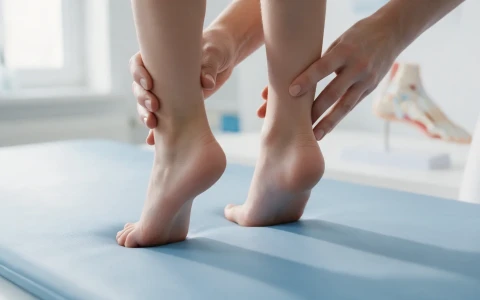
- A flattened arch that does not reappear even when standing on tiptoe
These symptoms are often put down to a simple sprain, a sports injury or growing pains. Pain is commonly felt on the outer side of the ankle, in the hollow just in front of the heel, or along the inner side of the foot. Repeated sprains on the same side, pain that lasts longer than expected after a sprain, or similar symptoms in both feet are clues that an underlying cause should be looked for.
Most flat feet in children are flexible and cause no symptoms. In tarsal coalition, however, the flat foot is rigid. The table below shows the difference; for a broader look at childhood foot and leg problems, see our page on childhood lower limb deformities.
| Feature | Flexible flat foot | Rigid flat foot (possible coalition) |
|---|---|---|
| Standing on tiptoe | Arch forms, heel turns inwards | No arch forms, heel stays straight |
| Hindfoot movement | Normal | Restricted |
| Symptoms | Usually none | Activity-related pain, recurrent sprains |
| General approach | Usually no treatment needed | Orthopaedic assessment and imaging needed |
How is tarsal coalition diagnosed?
Diagnosis begins with a careful examination. The doctor looks at the arch standing and on tiptoe, compares hindfoot movement on both sides and finds the tender areas. In some teenagers the muscles on the outer side of the leg tighten and hold the foot turned outwards.
The first imaging step is standing X-rays of the feet. An oblique view is particularly useful for showing a calcaneonavicular coalition, and on the side view a characteristic elongated bony projection may be seen. Indirect signs of a talocalcaneal coalition are looked for on the side view and on a special angled view of the heel. A CT scan is often requested to confirm the diagnosis, showing the size of the bridge, how much of the joint surface it involves and the state of the other foot. MRI is more useful for cartilaginous or fibrous coalitions and for associated findings such as bone marrow oedema. Imaging also helps rule out ankle ligament injuries and other causes of pain.
Non-surgical treatment options
For children and teenagers with symptoms, treatment usually starts without surgery:
- Temporarily pausing or reducing the activities that provoke pain
- Pain relief as advised by your doctor, and ice
- Insoles or orthoses that support the hindfoot
- A walking cast or walking boot for a few weeks when symptoms flare
- Physiotherapy for stretching, strengthening and balance, with an ankle support for sport
Symptoms settle for many young people with these measures, although in some they come back from time to time. The response varies from person to person.
When is surgery needed and what does it involve?
If pain persists despite non-surgical treatment and clearly limits daily life or sport, surgical options are considered. The choice depends on the type and size of the coalition, whether arthritis has developed, the shape of the arch and the patient's age.
- Resection of the bridge: the connection between the bones is removed and muscle or fat is placed in the gap to stop it re-forming. The aim is to reduce pain and restore some movement. It is generally preferred in younger patients without arthritis.
- Arch correction: if there is a marked flat foot, a corrective osteotomy of the heel bone may be planned in the same operation.
- Joint fusion (arthrodesis): for very large coalitions, established arthritis or when earlier treatment has not helped, the affected joints can be fused. This aims to reduce pain, but movement in those joints is lost.
| Type | Typical onset of symptoms | Imaging | Common surgery |
|---|---|---|---|
| Calcaneonavicular | Around 8–12 years | Often visible on an oblique foot X-ray | Resection with tissue interposition |
| Talocalcaneal | Around 12–16 years | CT or MRI usually needed | Resection; correction or fusion for large coalitions |
Recovery after surgery
After surgery, a cast or boot is usually worn for a few weeks, with no weight or only limited weight through the foot. After resection, starting movement exercises when your surgeon advises is important to keep the motion gained. Physiotherapy then works on strength, balance and walking. Returning to sport often takes a few months, and longer after a fusion, because the bones need to unite. Recovery depends on the type of operation and the individual.
When should you see a doctor?
- Repeated ankle sprains from minor twists
- Foot pain that increases with sport and does not fully settle with rest
- A rigid-looking flat foot in which the arch does not form on tiptoe
- Difficulty on uneven ground, or limping after sport
- Pain and swelling that continue for weeks after a sprain
- A teenager with a family history of tarsal coalition who develops foot symptoms
Early assessment allows non-surgical treatment to be used in a planned way and, if surgery is needed, for the decision to be made while the joints are still healthy. Our article on common paediatric deformities and early intervention and our paediatric orthopaedics page offer further reading. Prof. Dr. Halil İbrahim Balcı is a faculty member at Istanbul Faculty of Medicine working in paediatric orthopaedics and childhood lower limb deformities.
Key points
- Tarsal coalition is an abnormal bridge between foot bones, present from birth and often symptomatic in adolescence.
- Recurrent sprains, activity-related foot pain and a rigid flat foot are the main symptoms.
- Diagnosis relies on examination, standing foot X-rays and often CT or MRI.
- Treatment begins with insoles, a cast or boot, and physiotherapy.
- If symptoms persist, resection, corrective osteotomy or fusion may be considered; the decision is individual.
Frequently Asked Questions
Because the bridge between the bones is present from birth, it does not disappear by itself. Not everyone with a coalition has symptoms, however, and many teenagers who do find that their symptoms ease with non-surgical measures such as insoles, a short period in a cast or boot, and physiotherapy. Symptoms may come back from time to time. If pain persists and limits daily life or sport, surgical options are considered after examination and imaging.
In most cases, yes. Young people without symptoms, or whose symptoms are controlled with non-surgical treatment, can usually keep playing sport with suitable footwear, insoles and, if needed, an ankle support. During flare-ups it may be necessary to take a break or switch to less demanding activities for a while. Which sports are appropriate should be planned with your doctor, depending on the type of coalition, the severity of symptoms and the examination findings.
It depends on the type of operation. After resection of the bridge, a cast or boot is usually worn for a few weeks, followed by movement and strengthening work, and a gradual return to sport often happens within a few months. After a fusion, the bones need time to unite, so the return may take longer. The surgeon and physiotherapist decide the timing together, based on how recovery is progressing.
Tarsal coalition is known to occur in more than one member of some families, which suggests a genetic tendency. That does not mean a sibling or child of someone with a coalition will necessarily have one, and coalitions within a family do not always cause symptoms. If a child with a family history of tarsal coalition starts having repeated sprains or foot pain, an orthopaedic assessment is advisable.
Yes. In some people a coalition causes no symptoms during childhood and adolescence and is only discovered in adulthood, after a sprain, an increase in activity or imaging done for another reason. In adults, joints around a long-standing coalition may have developed arthritis, which affects the treatment options. Assessment involves examination, standing X-rays and, where needed, CT or MRI.
This article is for general information only and does not replace a medical examination, diagnosis or treatment. Treatment options and outcomes vary from person to person.
Related treatment area Childhood Lower Limb Deformities: Bow Legs, Knock Knees and In-Toeing