Bone transport is an Ilizarov technique that repairs large bone defects by generating new bone from the patient's own bone. How it works, the stages of treatment, techniques and what to expect.
On this page 8 sections
- What is bone transport? The principle of distraction osteogenesis
- When is bone transport used?
- The stages of treatment: from surgery to union
- Bone transport techniques
- How does bone transport compare with other reconstruction methods?
- Living with treatment and possible complications
- When should you contact your doctor?
- Key points
Bone transport is an Ilizarov technique for rebuilding large sections of bone lost through injury, infection or tumour, using the patient's own bone. The surgeon makes a controlled cut in healthy bone, and the freed segment is then moved into the gap at about 1 millimetre a day, using an external fixator or a special nail; the space left behind it gradually fills with new bone. In suitable patients, bone transport can repair defects measuring several centimetres, but it is a treatment lasting many months that requires patience and regular follow-up.
This article explains how bone transport works, when it is used, the stages and techniques of treatment, how it compares with other methods and what to look out for. The most appropriate method is chosen individually, according to the size and site of the bone loss, whether infection is present, the condition of the soft tissues and the patient's general health.
What is bone transport? The principle of distraction osteogenesis
The Soviet orthopaedic surgeon Gavriil Ilizarov showed that bone and the surrounding soft tissues can regenerate under slow, controlled tension. In this process, known as distraction osteogenesis, the ends of a carefully cut bone, with its blood supply preserved, are moved apart by a tiny amount each day. The body fills the widening gap with new bone tissue (the regenerate), which is soft at first and then gradually hardens.
The same principle is used in limb lengthening, but whereas lengthening increases the overall length of the bone, bone transport keeps the length the same and carries a segment of bone across a defect. As the transport segment advances, the gap in front of it shrinks and new bone forms behind it. In this way, bone loss is repaired with the patient's own bone tissue, without the need to harvest large amounts of bone from elsewhere.
When is bone transport used?
Bone transport is generally considered for bone loss of more than a few centimetres that would be difficult to repair by other means:
- bone loss after high-energy trauma and open fractures
- gaps left after removing infected bone in infected nonunion or chronic bone infection
- defects after removal of bone tumours, in selected patients
- complex nonunions combined with shortening and deformity
- certain childhood conditions, such as congenital pseudarthrosis of the tibia
The method can be used where infection has been cleared, and shortening and deformity can be corrected during the same treatment, so it is frequently used in managing complex and infected nonunion.
The stages of treatment: from surgery to union
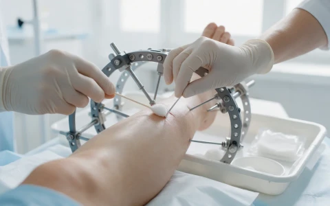
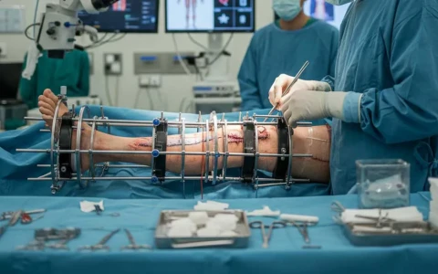
- Surgery: if there is infection, infected and non-viable bone and tissue are removed first. An external fixator (usually a circular Ilizarov frame or a monolateral rail system) is attached to the bone, and a controlled cut that preserves the blood supply (a corticotomy) is made in healthy bone away from the defect.
- Latency period: there is usually a wait of 5 to 10 days after surgery while healing begins at the corticotomy.
- Transport phase: the bone segment is moved by about 1 millimetre a day, usually split into three or four small adjustments. Patients and their families are taught how to make these adjustments. X-rays are typically taken every one to two weeks, and the rate is adjusted to the quality of the new bone and the patient's comfort.
- Docking: when the transport segment reaches the bone at the far end of the gap, the two ends need to unite. A small operation to freshen the bone ends, with a bone graft if needed, is frequently planned to help union at this docking site.
- Consolidation phase: the new bone left behind takes time to harden and strengthen, and this phase often lasts longer than the transport itself. As a rough guide, a total frame time of one to two months is allowed for each centimetre transported, although this varies considerably from person to person.
- Frame removal and rehabilitation: once X-rays show that the new bone is strong enough, the fixator is removed. Some patients need a period of protection in a cast or brace, followed by physiotherapy.
Bone transport techniques
Bone transport is not a single operation but a family of techniques tailored to each patient:
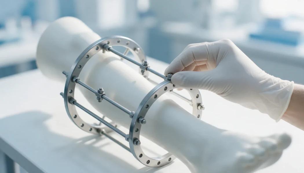
- Circular (Ilizarov) frame transport: rings attached to the bone with wires and pins give stable fixation, allow most patients to walk and bear weight in the frame, and make it possible to correct deformity at the same time.
- Monolateral rail fixators: less bulky, and may be preferred particularly in the thigh bone.
- Bifocal and trifocal transport: one segment can be moved through a single cut (bifocal), or two segments through two separate cuts (trifocal). In trifocal transport the two segments travel towards each other, which can help to shorten treatment for large defects.
- Transport over a nail and combined methods: transport over an intramedullary nail can shorten frame time, although the risk of infection is weighed carefully.
- Acute shortening and relengthening: for smaller defects, the gap is closed immediately by bringing the bone ends together, and the resulting shortening is later corrected by lengthening at another level.
- Internal transport systems: in selected patients, transport can be performed with special nails or cables placed inside the bone.
For an overview of where the Ilizarov method is used more generally, see our article What is Ilizarov surgery?
How does bone transport compare with other reconstruction methods?
Each option for large bone defects has its own advantages and limitations:
| Method | How it works | Key features |
|---|---|---|
| Bone transport | New bone is generated gradually from the patient's own bone | Suitable for large defects and after infection; shortening and deformity can be corrected at the same time; long frame time |
| Two-stage induced membrane technique | A biological membrane is first created around bone cement, and the gap is later filled with bone graft | May not need a frame; requires a large amount of graft and at least two operations |
| Vascularised bone transfer | A piece of bone (for example the fibula) is moved with its own blood vessels into the defect | Requires microsurgery; the transferred bone thickens over time |
| Acute shortening | The gap is closed straight away by bringing the bone ends together | Suitable for smaller defects; limited by the soft tissues; later lengthening may be needed |
These methods are sometimes combined, and the choice is made after examination, imaging and, where needed, joint assessment with plastic surgeons.
Living with treatment and possible complications
Bone transport requires active involvement from patients and their families. Cleaning the pin and wire sites regularly as advised, making frame adjustments on time, bearing weight as far as the surgeon allows and keeping up physiotherapy to prevent stiffness in neighbouring joints such as the knee and ankle are all essential. The long treatment may require adjustments at school, work and home, and psychological support can help.
The main problems that can arise during treatment include:
- infection at pin and wire sites (the most common problem, usually controlled with pin care and oral antibiotics)
- pain and tightness of the soft tissues during transport
- joint stiffness and muscle contractures
- delayed union or soft tissue becoming trapped at the docking site
- the new bone failing to strengthen adequately, or hardening too early
- the transport segment drifting out of line
- a fracture through the new bone after the frame is removed
Most of these can be recognised early and managed with regular follow-up. For the wider challenges of limb reconstruction, see our article on challenges in limb reconstruction surgery.
When should you contact your doctor?
During bone transport treatment, contact your team without waiting for your next appointment if you notice:
- increasing redness, swelling, discharge or pain at the pin or wire sites
- fever or shivering
- new numbness, tingling or weakness in the foot or hand
- severe pain after an adjustment that does not settle
- a loose or broken wire, pin or part of the frame
- increasing difficulty straightening the knee or lifting the foot at the ankle
- sudden pain, swelling or a change in shape after the frame has been removed
- pain and swelling in the calf, shortness of breath or chest pain
Bone transport is part of Ilizarov and limb reconstruction surgery and calls for detailed planning and experience. Prof. Dr. Halil İbrahim Balcı is a co-editor of the Springer book on external fixator applications according to Ilizarov principles, published in 2018, and worked as a research fellow at the Paley Institute on deformity surgery and Ilizarov techniques.
Key points
- Bone transport is an Ilizarov technique that repairs large bone defects by gradually generating new bone from the patient's own bone.
- A cut segment of bone is moved about 1 millimetre a day, and the gap left behind it fills with new bone.
- Treatment passes through latency, transport, docking and consolidation phases and usually lasts many months.
- Pin site care, physiotherapy and regular X-ray follow-up are essential parts of treatment.
- The choice between bone transport and other reconstruction methods is made individually after examination and imaging.
Frequently Asked Questions
The length of treatment depends mainly on how much bone needs to be transported. Transport proceeds at about 1 millimetre a day, followed by a period in which the new bone hardens. As a rough guide, a total frame time of one to two months is allowed for each centimetre transported, but age, smoking, a history of infection and healing at the docking site can all change this. Rehabilitation continues for some time after the frame is removed.
Pain and tightness can be felt in the first few days after surgery and during the transport phase, particularly after adjustments. The level of discomfort varies from person to person and can usually be managed with appropriate pain relief, physiotherapy and, if needed, by adjusting the rate of transport. Increasing pain at a pin site can be a sign of infection and should be reported to the team. It is worth discussing a pain management plan with your doctor before treatment starts.
Most patients, particularly those with circular frames, can walk with crutches and put weight through the leg as far as the surgeon allows, and this can also encourage new bone formation. How much weight you can bear, however, depends on where the bone loss is, the type of fixator, whether there is infection and the stage of treatment. Your weight-bearing programme is set at your post-operative reviews and updated as treatment progresses.
Bone transport generates new bone gradually from the patient's own bone and usually requires a frame for a long period. In the Masquelet (two-stage induced membrane) technique, bone cement is first placed in the gap so that a biological membrane forms around it; several weeks later the cement is removed and the space is filled with bone graft. Each method has its own advantages, and the choice depends on the size of the defect and the patient's circumstances.
Yes, in suitable cases. Children generally have a strong capacity to regenerate bone, but treatment must be planned carefully to protect the growth plates, allow for future growth and take account of the effect on the child's schooling and social life. Congenital pseudarthrosis of the tibia and bone loss after trauma or infection are among the situations in which the method may be used in this age group.
This article is for general information only and does not replace a medical examination, diagnosis or treatment. Treatment options and outcomes vary from person to person.
Related treatment area Revision Surgery for Complex and Infected Nonunion