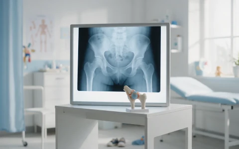
Coxa vara is a reduced angle between the neck and shaft of the thighbone, which can cause a limp and a leg length difference in children. Types, diagnosis and treatment options.
On this page 8 sections
Coxa vara is a condition in which the angle between the neck of the thighbone (femur), which leads up to the hip, and the shaft of the bone is smaller than normal. When this angle is reduced, the muscles around the hip work at a mechanical disadvantage; children usually present with a limp that does not typically hurt, a shorter leg on the affected side or, when both hips are involved, a waddling walk. Coxa vara may be present from birth, develop during growth or result from another condition. Depending on the size of the angle, whether it is progressing and the symptoms, treatment ranges from observation to surgical realignment of the upper end of the thighbone.
Coxa vara is rare, and for most families it is an unfamiliar term. This article explains what the angle means, the possible causes, the measurements used in diagnosis and the treatment options in plain language.
What is coxa vara? Understanding the hip angle
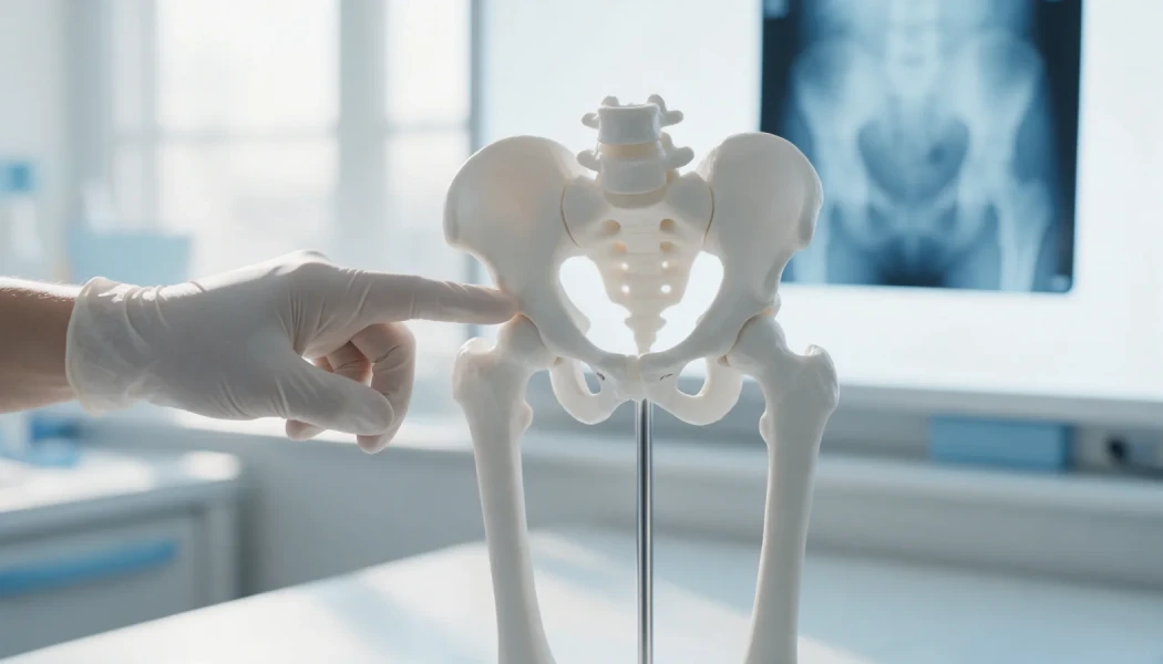
At the top of the thighbone, the ball-shaped femoral head that sits in the hip socket is joined to the shaft of the bone by an angled "neck". The angle between the neck and the shaft is called the neck-shaft angle. It is wider at birth and gradually decreases with growth, reaching roughly 125-135 degrees in adults.
In children, a marked reduction in this angle, usually to below 120 degrees, is defined as coxa vara. As the angle becomes smaller, the femoral head tilts downwards, the bony prominence on the side of the hip known as the greater trochanter rides higher, and the muscles that move the leg out to the side work less effectively. The opposite, an angle larger than normal, is called coxa valga.
The angle matters because of the way the hip abductor muscles work. These muscles run from the greater trochanter to the pelvis and keep the pelvis level when you stand on one leg. When the trochanter moves upwards, the muscles slacken and have to work through a shorter lever arm; as a result, the pelvis drops towards the opposite side during walking and a limp appears. With marked or progressive deformity, the extra stress on the femoral neck makes careful monitoring all the more important.
Types and causes of coxa vara
Coxa vara is not a single disease but a deformity that can arise for different reasons. Doctors generally consider three main groups:
| Type | When is it noticed? | Features |
|---|---|---|
| Congenital coxa vara | At birth or in infancy | Often seen together with congenital shortening or deficiency of the femur |
| Developmental coxa vara | After a child starts walking, usually in the preschool years | Caused by abnormal bone formation in the lower part of the growth plate in the femoral neck; may affect both hips |
| Acquired coxa vara | At any age | Linked to conditions such as hip fractures, hip infections, slipped capital femoral epiphysis, Perthes disease, skeletal dysplasias, rickets or fibrous dysplasia |
In developmental coxa vara the growth plate in the femoral neck is more vertical than normal, and a triangular fragment of bone may be seen on X-ray at the lower part of the neck. Because body weight acts on this steep growth plate as a shearing force, the angle can become smaller over time. For other rare conditions in this group, see our article on rare childhood deformities.
What are the signs of coxa vara?
Most children with coxa vara do not have significant hip pain; parents tend to notice a difference in the way their child walks. Common findings include:
- A limp, with the upper body leaning towards the affected side when walking
- A waddling, side-to-side walk when both hips are affected
- A difference in leg length when only one side is affected
- Reduced ability to move the leg out to the side and to turn the hip inwards
- Tiring quickly after long walks or running
- A prominent bony bump on the side of the hip
Because a leg length difference can affect walking on its own, its size and how it is likely to change with growth are also assessed during follow-up. Our article on leg length discrepancy in children covers this in detail.
Diagnosis: X-rays and angle measurements
At the examination, the doctor assesses walking, hip movement, leg lengths and the strength of the muscles that move the hip out to the side. When the pelvis drops towards the opposite side as the child stands on one leg (the Trendelenburg sign), it shows that these muscles are not working effectively.
The diagnosis is made with X-rays of the pelvis and hips, on which the following are measured:
- Neck-shaft angle: shows how severe the deformity is.
- Hilgenreiner-epiphyseal (HE) angle: shows how steeply the growth plate in the femoral neck is tilted, and helps predict whether the deformity is likely to progress.
In children with suspected congenital or acquired coxa vara, full-length X-rays of the legs, CT or MRI where needed, and further tests if a skeletal dysplasia is suspected may also be requested. In particular, for children with coxa vara on both sides who are noticeably shorter than their peers, a wider assessment of the skeleton and, where appropriate, genetic testing may be suggested to look for a generalised disorder of bone development.
Treatment of coxa vara: observation or surgery?
Treatment decisions take into account the measured angles, whether the deformity is progressing over time, how much walking is affected and the child's age. In developmental coxa vara, a general guide is:
- With an HE angle below about 45 degrees, the deformity often does not progress and is monitored with regular X-rays.
- With angles between 45 and 60 degrees, the child is followed closely, and surgery is considered if the deformity progresses or causes symptoms.
- With angles above 60 degrees, or a markedly reduced neck-shaft angle, the deformity is more likely to progress, so surgery is more often recommended.
Physiotherapy and exercise can help strengthen the muscles around the hip and support walking, but they do not change the shape of the bone. In acquired coxa vara, treatment is planned with the underlying condition in mind.
Valgus osteotomy and recovery after surgery
The operation commonly used for coxa vara is a valgus osteotomy, in which a controlled cut is made at the upper end of the thighbone to increase the neck-shaft angle. This brings the femoral neck to a more favourable angle, repositions the growth plate so that it is compressed rather than sheared by body weight, and improves the working conditions of the muscles that move the hip out to the side. The bone is usually held with a plate and screws. Achieving the planned angle is important in reducing the risk of the deformity recurring.
Before a decision is made, the following are usually discussed in detail with the family:
- The aim of surgery and the planned correction
- The hospital stay, the period in a cast or on crutches, and returning to school
- Possible risks such as infection, delayed bone healing and recurrence of the deformity
- Whether the plate and screws may need to be removed later
- The planned schedule of check-ups during growth
Recovery depends on the child's age and the procedure performed. Young children may spend a period in a cast; older children usually go through a phase of protected weight-bearing on crutches. Once the bone has healed, physiotherapy helps to rebuild muscle strength and walking.
Follow-up continues until growth is complete. In some children the growth plate in the femoral neck closes early, the greater trochanter becomes relatively high or a leg length difference develops; further procedures may then be planned. When coxa vara accompanies congenital shortening of the femur, hip correction is part of a wider reconstruction plan that may also include stages such as bone lengthening later on.
When should you see a doctor?
An assessment by a paediatric orthopaedic specialist is advisable if your child has:
- A limp that persists or becomes more obvious after they start walking
- A waddling, side-to-side walk
- A difference in leg length, or shoe soles that wear unevenly
- Reduced hip movement, particularly when moving the leg out to the side
- A new change in walking after a previous hip fracture or hip infection
- Hip or walking problems alongside a diagnosed skeletal dysplasia
Prof. Dr. Halil İbrahim Balcı worked as a research fellow at the Paley Institute, with a focus on congenital bone anomalies and paediatric orthopaedics. Coxa vara is assessed within rare childhood limb deformities and paediatric orthopaedics, with treatment tailored to each child's examination and X-ray findings.
Key points
- Coxa vara means the angle between the neck and the shaft of the thighbone is smaller than normal.
- It may be congenital, developmental or acquired as a result of other conditions.
- It is usually noticed as a limp that does not typically hurt, a waddling walk or a leg length difference.
- The neck-shaft angle and the HE angle measured on X-ray guide the choice between observation and surgery.
- For progressive or marked deformity a valgus osteotomy may be performed, and follow-up until growth is complete is important.
Frequently Asked Questions
In developmental coxa vara, if the growth plate is not tilted too steeply, the deformity often does not progress and some children improve over time. Where the growth plate is markedly vertical, however, the angle may become smaller as the child grows. Which group a child falls into is determined by the HE angle measured on X-ray and by regular check-ups, and the course varies from child to child.
There is no single ideal age for surgery. The decision depends on the size of the angle, whether the deformity is progressing, how walking is affected and the child's bone development. In developmental coxa vara, surgery is often considered in the preschool or early school years. When coxa vara accompanies congenital shortening of the femur, timing is decided as part of the overall reconstruction plan.
In one-sided coxa vara the affected leg may be shorter because the femoral neck tilts downwards. When coxa vara accompanies congenital shortening of the femur the difference is more marked and may increase with growth. Early closure of the growth plate after surgery can also create a difference. Leg lengths are therefore measured regularly during follow-up, and methods to balance any difference are planned if needed.
No. In developmental dysplasia of the hip, the main problem is a shallow socket in which the femoral head does not sit fully. In coxa vara the femoral head usually remains in the socket; the problem is the reduced angle between the neck and the shaft of the thighbone. As both can cause a limp, they are distinguished by examination and X-ray.
Physiotherapy and exercise can help strengthen the muscles that move the hip out to the side, supporting walking and reducing fatigue. They also play an important part in rebuilding strength after surgery. However, they do not change the angle of the bone. Where the angle is marked or progressing, surgical options are considered to correct the shape of the bone.
This article is for general information only and does not replace a medical examination, diagnosis or treatment. Treatment options and outcomes vary from person to person.
Related treatment area Rare Childhood Limb Deformities: Fibular Hemimelia, Femoral Deficiency