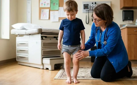
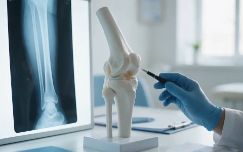
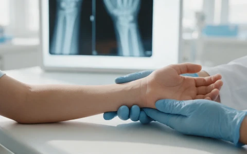
Rickets can soften children's bones and cause bowed legs, most often because of vitamin D deficiency. We explain the signs, how it differs from normal bowing, and how it is diagnosed, treated and prevented.
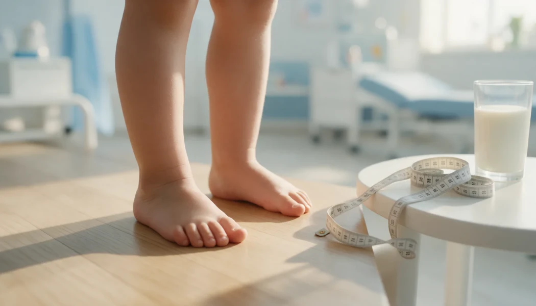
On this page 8 sections
Rickets is a softening and bending of the bones in growing children caused by poor mineralisation, and its most common cause is vitamin D deficiency. The weight-bearing leg bones are affected most, which is why rickets is often first noticed as bowed legs or knock knees in a toddler who has recently started walking. When rickets and bowed legs are recognised early and treated, the bones start to mineralise normally again, and in young children much of the bowing can correct with growth.
This article explains what causes rickets, how it differs from the normal bowing seen in toddlers, how it is diagnosed and treated, and when orthopaedic surgery may be considered. Every child is different, and the right approach is decided after examination, blood tests and imaging.
What is rickets and what causes it?
For bone to harden, calcium and phosphate have to be laid down in the bone tissue. Vitamin D acts like a hormone that allows the gut to absorb these minerals. When vitamin D, calcium or phosphate is lacking, the cartilage in the growth plates cannot turn into bone properly: the growth plates widen, the bone softens and it bends under body weight. In adults, whose growth plates have closed, the same problem causes bone pain and fragility rather than bowing, and is called osteomalacia.
Types of rickets
- Nutritional rickets: the most common type, caused by too little sunlight, missing vitamin D supplements or a diet low in calcium.
- Hypophosphataemic rickets: a mostly inherited condition in which the kidneys lose phosphate. Bowing develops even though vitamin D levels are normal, so it is also known as "vitamin D-resistant rickets".
- Other causes: kidney and liver disease, malabsorption such as coeliac disease, and rare genetic conditions that affect how vitamin D is activated in the body.
Risk factors
- babies who are exclusively breastfed without a vitamin D supplement (breast milk is an excellent food, but it contains little vitamin D);
- low maternal vitamin D during pregnancy, and premature birth;
- darker skin, covered clothing and spending very little time outdoors;
- winter months, when sunlight is too weak for the skin to make enough vitamin D;
- a diet low in calcium, with little milk or dairy;
- chronic illness and long-term use of certain medicines that affect vitamin D metabolism.
Signs of rickets: bowed legs and other clues
Symptoms usually appear between about 6 months and 3 years of age, during a period of rapid growth. Leg bowing is the most visible sign, but rarely the only one:
- bowed legs or knock knees that get worse after the child starts walking;
- thickening and widening of the wrists and ankles;
- bead-like swellings where the ribs meet the breastbone (the "rachitic rosary");
- late closure of the soft spot on the head, softening of the skull bones and a prominent forehead;
- delayed teething and defects in tooth enamel;
- muscle weakness, late sitting or walking, a waddling gait and bone pain;
- slow growth in height and fractures that happen easily.
Babies whose calcium level falls very low can develop muscle spasms or seizures, which need urgent medical attention.
Normal bowing or rickets?
Bowed legs are not always a sign of disease. Most babies are born with bowed legs; this usually straightens by around 18–24 months, a mild knock-knee phase follows at about 3–4 years, and by around 7–8 years most children's legs are close to adult alignment. The table compares three common situations:
| Feature | Physiological (normal) bowing | Bowing due to rickets | Blount disease |
|---|---|---|---|
| Age | Bowing up to 2 years, mild knock knees at 3–4 years | Usually after the child starts walking | Often 2–4 years in early walkers; can also appear in adolescence |
| Symmetry | Both sides similar | Usually both sides | May be one-sided or asymmetrical |
| Course | Corrects on its own over time | May persist or worsen without treatment | Usually progressive |
| Other findings | None; height and development normal | Thick wrists, delayed teeth, short stature | Sharp angulation at the top of the shin bone |
| Blood tests | Normal | Usually abnormal | Usually normal |
For normal stages of leg development and when to monitor, see our parents' guide to leg bowing; for a growth-plate condition of the shin bone that is an important alternative diagnosis, read our article on Blount disease.
How is rickets diagnosed?
The diagnosis combines the child's history, a clinical examination, blood tests and X-rays. The doctor will ask about diet, vitamin D supplements, time spent outdoors and any family history of bowed legs or short stature.
- Blood tests: calcium, phosphate, alkaline phosphatase (ALP), 25-hydroxyvitamin D and parathyroid hormone (PTH), with kidney function and urinary phosphate where needed. The pattern of results helps to distinguish nutritional rickets from hypophosphataemic rickets.
- X-rays: an X-ray of the wrist or knee typically shows widened growth plates and bone ends that are cupped and frayed. A standing full-length X-ray of the legs shows how much bowing there is and where it lies.
Treating rickets: vitamin D, calcium and orthopaedic surgery
Correcting the deficiency
The first and most important step is to correct the underlying deficiency. In nutritional rickets, vitamin D and, when needed, calcium are given at a dose and for a period set by the paediatrician or paediatric endocrinologist, and progress is checked with blood tests and X-rays. High doses of vitamin D taken without medical advice can be harmful, so dosing should always be guided by a doctor. Hypophosphataemic rickets needs a different treatment, planned by specialist teams.
Will the bowed legs straighten?
In young children, once the deficiency is treated, much of the mild to moderate bowing can improve noticeably as the child grows. This can take from several months to a few years, and the degree of improvement varies from child to child. There is no strong evidence that splints or special shoes correct the bowing caused by rickets.
When is orthopaedic surgery needed?
Orthopaedic treatment is considered when the bowing persists or progresses despite good metabolic control, or causes pain or walking problems:
- Guided growth: in children whose growth plates are still open, small plates temporarily slow growth on one side of the growth plate so that the leg straightens as it grows. It is done through small incisions.
- Osteotomy: in children nearing the end of growth, teenagers and adults, the bone is cut in a controlled way and realigned, either with internal fixation such as plates and screws or gradually with an external fixator.
Timing matters: if correction is carried out before the metabolic problem is under control, the deformity is more likely to recur. Orthopaedic surgeons therefore work closely with paediatricians and paediatric endocrinologists. You can read more on our page about childhood lower limb deformities.
Preventing rickets: sunlight, diet and vitamin D supplements
Nutritional rickets is largely preventable, and early summer is a good time to review sunlight and vitamin D:
- Supplements in infancy: many countries recommend a vitamin D supplement for babies, especially breastfed babies. In Turkey, the Ministry of Health programme offers vitamin D to infants from the first weeks of life, and there is also a programme for pregnant women. Your child's doctor will advise on the dose and duration.
- Sunlight: the skin makes vitamin D in response to UVB rays, which window glass largely blocks. How much time is needed depends on skin tone, season and time of day. Sunburn should always be avoided, and babies under 6 months should be kept out of direct sun; at this age sunlight is not a substitute for supplements.
- Diet: milk, yoghurt and cheese are rich in calcium. Egg yolk and oily fish such as anchovies, sardines and salmon contain vitamin D, although food alone is often not enough.
- Higher-risk children: children who wear covering clothes, have darker skin, spend long periods indoors or have a chronic illness should have their vitamin D status reviewed more closely by their doctor.
When should you see a doctor?
It is worth having your child assessed by a paediatrician and, where needed, a paediatric orthopaedic surgeon if you notice:
- bowing that increases after the age of 2 or persists beyond 3;
- knock knees that are marked for the child's age or persist beyond 7–8 years;
- bowing on one side only, or a clear difference between the two legs;
- slowing growth in height, or thickened wrists and ankles;
- late walking, tiring easily, bone pain or frequent falls;
- a family history of bowed legs, short stature or rickets.
Muscle spasms, twitching or seizures in a baby need emergency care. The orthopaedic follow-up of bowing caused by rickets falls within paediatric orthopaedics, and Prof. Dr. Halil İbrahim Balcı assesses and treats leg deformities in children.
Key points
- Rickets is poor mineralisation of growing bones, most often caused by vitamin D deficiency.
- Bowed legs are often accompanied by thick wrists, delayed teething and slow growth.
- Diagnosis relies on blood tests and X-rays, and treatment starts with correcting the deficiency.
- In young children bowing can improve considerably with treatment; persistent deformity may need guided growth or osteotomy.
- Vitamin D supplements in infancy, safe sun exposure and a calcium-rich diet are the foundations of prevention.
Frequently Asked Questions
Not while the deficiency remains untreated, because soft bone keeps bending under load. Once vitamin D and calcium deficiency have been corrected under medical supervision, much of the mild to moderate bowing in young children can lessen with growth. Improvement may take from several months to a few years. Regular standing X-rays of the legs during this time show whether the alignment is improving or whether surgery needs to be considered.
Vitamin D status is measured with a blood test for 25-hydroxyvitamin D. When rickets is suspected, this test alone is not enough: calcium, phosphate, alkaline phosphatase and parathyroid hormone are checked as well. Looking at these results together shows whether the deficiency is affecting the bones and helps identify the type of rickets. A wrist or knee X-ray may also be requested if needed.
Sunlight is the main natural source of vitamin D, but whether it is enough depends on season, time of day, skin tone, clothing and time spent outside. In winter and behind glass the skin makes very little. For babies, sunshine does not replace a vitamin D supplement, and infants under 6 months should be kept out of direct sun. Sunburn should be avoided at every age; for children at higher risk, a doctor may recommend a supplement based on blood levels.
Nutritional rickets is caused by a lack of vitamin D or calcium and is expected to improve once the deficiency is corrected. Hypophosphataemic rickets is a mostly inherited condition in which the kidneys lose too much phosphate; vitamin D levels may be normal and the condition does not respond to vitamin D alone. A family history of bowing, short stature and bowing that persists despite treatment point towards it. Treatment is planned with paediatric endocrinology and nephrology teams.
There is no fixed age; the decision depends on how severe the bowing is, how well the metabolic problem is controlled and whether the growth plates are still open. In growing children with persistent bowing despite medical treatment, guided growth may be considered. In children near the end of growth and in adults, an osteotomy may be discussed. In every case the metabolic problem should be under control first, and timing is decided individually after examination and imaging.
This article is for general information only and does not replace a medical examination, diagnosis or treatment. Treatment options and outcomes vary from person to person.
Related treatment area Childhood Lower Limb Deformities: Bow Legs, Knock Knees and In-Toeing