Noticing asymmetry or deformity during your child's physical development can understandably raise concerns. Especially in toddlers who are just beginning to walk, the alignment of the legs may sometimes appear atypical. While leg bowing in children is often a natural part of growth and development, certain cases may require medical evaluation and intervention.
This guide covers the common causes of leg bowing in growing children, outlines what is considered within normal limits, and explains when treatment may be necessary. Our goal is to help parents monitor their child's growth knowledgeably and seek expert advice at the right time.
What Is Leg Bowing in Children?
Leg bowing in children occurs when the angle formed between the thigh bone (femur) and shinbone (tibia) at the knee joint deviates from the normal anatomical axis. This results in the legs appearing curved outward (bowlegs or genu varum) or inward so that the knees touch (knock-knees or genu valgum). As growth plates remain active throughout childhood, the shape and angle of the bones dynamically change with age.
Most deviations arise temporarily due to fetal positioning or the distribution of weight when a child begins to walk. However, bone metabolism disorders, trauma, or developmental abnormalities can cause permanent deformities.
Distinguishing Physiological vs. Pathological Leg Bowing
When evaluating leg bowing, the key distinction is whether the condition is "physiological" (a normal part of development) or "pathological" (caused by disease). Physiological bowing is usually symmetrical, painless, and corrects naturally with growth. Pathological cases may worsen over time and require treatment.
For more detailed information, visit our page on childhood lower limb deformities.
Physiological Bowlegs (Genu Varum)
Infants are generally born with slight bowlegs due to their position inside the womb. This condition is considered normal up to about 2 years of age when evaluating leg bowing in children. As the child starts walking and leg muscles strengthen, this bowing typically improves between 18 and 24 months, eventually reaching a neutral alignment. In physiological bowlegs, when the child stands, the ankles touch while the knees stay apart with a small but not excessive gap that decreases over time.
Physiological Knock-Knees (Genu Valgum)
After age 2, children's leg alignment often shifts from bowlegs toward knock-knees, peaking around ages 3 to 4. During this phase, the knees touch while the ankles are spaced apart. By age 7 to 8, the legs usually assume a typical adult alignment.
Pathological Deformities and Warning Signs
If the bowing exceeds normal age limits or if warning signs appear, the condition might be pathological. Diseases such as Blount’s disease, rickets (vitamin D deficiency), skeletal dysplasias, or growth plate injuries following trauma can cause pathological bowing.
Particularly in rare childhood deformities described on our rare pediatric deformities page, the following signs should be closely monitored:
- Bowing affecting only one leg (asymmetry).
- Short stature compared to peers (growth retardation).
- Pain, limping, or early fatigue during walking.
- Progressive worsening of bowing instead of natural improvement.
When Should Leg Bowing Be a Concern?
For parents, the most challenging aspect is knowing when to move beyond a "wait-and-see" approach and seek medical care. In general, any leg alignment inconsistent with the child’s age warrants a specialist consultation. For example, severe bowlegs at age 3 or pronounced knock-knees at age 10 are abnormal.
Moreover, commonly seen pediatric deformities identified early respond better to simple treatments. Excessive knee angulation can lead to premature cartilage wear (osteoarthritis) and meniscus problems later if neglected.
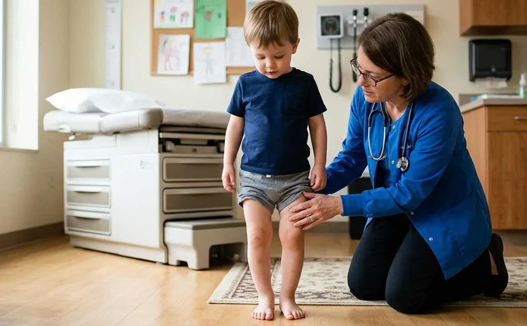
Diagnostic Process: What Tests Are Performed?
Diagnosis begins with a thorough physical examination. The physician will assess the child's gait (walking pattern), joint range of motion, and leg lengths. Weight-bearing full-length leg X-rays ("orthoroentgenograms") are the most commonly used imaging method to measure the degree of bowing. Mechanical axes are drawn through the centers of the hip, knee, and ankle joints to quantify deviation.
If a metabolic bone disorder is suspected (e.g., rickets), blood tests measuring calcium, phosphate, alkaline phosphatase, and vitamin D levels are performed. In certain cases, genetic screening or MRI scans are also warranted to evaluate bone development disorders in children.
Treatment Methods: When and What Is Recommended?
Treatment plans depend on the cause, severity, and age of the child. For general information on treatment options, please see our article on O-Leg and X-Leg treatments.
Observation and Watchful Waiting
Mild physiological bowing usually does not require treatment. The child is monitored clinically and radiologically every 3 to 6 months. Parents should support good nutrition and keep up with doctor appointments. Most physiological bowing spontaneously corrects during this time.
Orthoses and Physical Therapy
In early-stage Blount’s disease or some metabolic conditions, braces (orthoses) can help stop progression and correct bowing by balancing load on the growth plates and encouraging proper bone growth. Physical therapy supports muscle balance and joint mobility.
Surgical Intervention
Surgery is considered when conservative management (observation, medication, orthoses) fails in advanced deformities, persistent bowing during adolescence, or pathological bone diseases. Surgical options include growth modulation (hemiepiphysiodesis or “8-plate” technique) and corrective osteotomy (bone cutting and realignment).
In cases of improperly healed fractures, malunion correction surgery is performed to restore leg axis. Complex reconstructions may also be necessary for failed prior surgeries or infection-related complications, combining nonunion surgery and lower limb deformity correction principles. Growth modulation surgery is minimally invasive, temporarily arresting the growth plate to allow natural correction through the child’s own growth potential.
Simple Home Observations
Parents can monitor leg development at home through simple checks. Have your child stand in shorts on a flat surface with feet together, facing forward. Observe the distance between their knees and ankles. Watch for knees knocking together while walking, or feet turning inward or outward, as these are useful clues.
Summary of Home Monitoring
Leg bowing in children is often a temporary phase of growth. However, attentive parental observation and timely consultation with a pediatric orthopaedist help prevent permanent issues. Early diagnosis is the most important factor for successful treatment.
This content is provided for general informational purposes only and is not intended as professional medical advice, diagnosis, or treatment. If you have concerns about your child's health, please consult a qualified healthcare professional for proper diagnosis and treatment. The information on this site should not be used to create a personal treatment plan without a medical examination. Do not delay seeking medical care based on the content provided.