Does your MRI report mention bone marrow oedema? We explain in plain language what the finding means, its common causes, the terms used in reports, treatment options and how long recovery takes.
On this page 8 sections
Bone marrow oedema is an MRI finding that shows increased fluid inside a bone. It is not a disease in itself but a sign that the bone is reacting to load, injury or an underlying problem, so treatment depends on what is causing it. American reports spell it "edema", so you may see the phrase bone marrow edema on your scan report. Most cases caused by an impact or overload settle over weeks to months once the load is reduced, while others need the underlying joint or bone problem to be treated as well.
Many people who read "bone marrow oedema" on their MRI report wonder what it means and how serious it is. This article explains the finding, its common causes, the terms used in reports and the treatment options. The right interpretation is only possible when the images are considered alongside your symptoms and examination.
What is bone marrow oedema and why does it show on MRI?
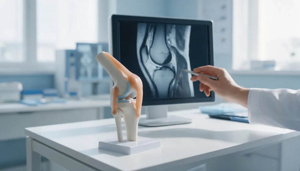
Inside our bones is the bone marrow, a spongy tissue rich in blood vessels and fat. When a bone is exposed to an impact, repetitive load, a problem with its blood supply or inflammation, the amount of fluid in this area increases. On MRI sequences that make fluid look bright (STIR or fat-suppressed T2 images) the area appears white, while on T1 images it looks dark.
Bone marrow oedema does not show on plain X-rays and is usually not visible on CT either, which is why it often comes to light on MRI in people whose X-rays are normal but whose pain persists. The "oedema" may not be just fluid: tiny fractures, small bleeds and repair tissue can create the same appearance. For this reason radiologists sometimes use the phrase "oedema-like signal change".
What causes bone marrow oedema (edema)?
The same MRI finding can have very different causes. The main ones are:
- Impacts and sprains (bone bruise): common after knee sprains, anterior cruciate ligament injuries, kneecap dislocations and ankle sprains, at the points where bones knock against each other.
- Overload (stress reaction): develops from repetitive loading in running and jumping sports and can be the early stage of a stress fracture.
- Osteoarthritis: oedema can appear in the bone beneath areas of thinned cartilage, related to overload, and may be linked to pain.
- Subchondral insufficiency fracture: a small fracture just beneath the joint surface, most often in the knee of older adults, that appears without a clear injury and may accompany a meniscal root tear.
- Transient bone marrow oedema syndrome: seen mostly in the hip, in middle-aged men and in women late in pregnancy. Its cause is not fully understood and it usually settles on its own over several months.
- Avascular necrosis: loss of blood supply to part of a bone, which may first show as bone marrow oedema. Long-term steroid use and heavy alcohol intake are among the risk factors.
- Less common causes: bone infection, inflammatory arthritis, complex regional pain syndrome and bone tumours.
What do the terms in an MRI report mean?
MRI reports are written in technical language. The table translates some of the phrases that often appear alongside bone marrow oedema:
| Phrase in the report | What it means |
|---|---|
| Bone marrow oedema / oedema-like signal | Fluid and tissue reaction within the bone; the cause is interpreted together with clinical information |
| High signal on STIR or fat-suppressed T2 | Brightness consistent with oedema on the sequences that highlight fluid |
| Bone bruise (contusion) | A "bruise" inside the bone caused by an impact or twist |
| Subchondral | The bone directly beneath the joint cartilage |
| Stress reaction | Overload damage to the bone without a clear fracture line yet |
| Insufficiency fracture | A small fracture in weakened bone caused by everyday loads |
| Osteonecrosis / avascular necrosis | Loss of blood supply to part of the bone |
The report will also describe the location and size of the oedema and any associated findings, such as cartilage damage, a ligament or meniscal tear, or a fracture line. The word "oedema" on its own says little about severity; it is the full picture that guides the assessment.
Symptoms of bone marrow oedema
Symptoms vary with the cause and the location. Common complaints include:
- a deep, aching pain that worsens with weight-bearing, walking or using stairs;
- pain at rest and at night;
- swelling around the joint, stiffness and a limp;
- tenderness when pressing over the bone.
The site of the pain usually matches the site of the oedema: the inner or outer side of the knee, the groin for the hip, or deep within the ankle. Oedema in the hip can sometimes cause pain that spreads to the front of the thigh and the knee.
In some people, bone marrow oedema is found by chance on an MRI performed for another reason and causes no symptoms at all. That is why its significance must always be judged together with symptoms and examination findings.
Treatment of bone marrow oedema
The basic principle is to deal with the cause and give the bone time to recover. Treatment is tailored to the individual after examination and imaging, but general approaches include:
Reducing load and supportive care
- Offloading: walking with crutches and partial weight-bearing during the painful phase, and pausing activities that aggravate the pain. In transient bone marrow oedema of the hip, this also helps to reduce the risk of fracture while the bone is weakened.
- Physiotherapy: low-impact exercises to maintain joint movement and muscle strength.
- Bone health checks: vitamin D, calcium and, where needed, bone density are assessed, and any deficiency is treated under medical supervision.
- Weight management: reduces the load on the joint when oedema is related to knee osteoarthritis.
- Pain relief and other options: painkillers are used as advised by your doctor. Certain medicines and shockwave therapy are used in selected patients, although the evidence for them is limited.
When is surgery considered?
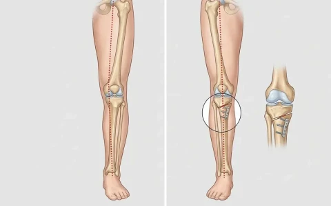
If the oedema is part of an underlying structural problem, that problem may need treating. For example, arthroscopic surgery may be considered for an associated meniscal root tear or cartilage damage, and realignment surgery such as a high tibial osteotomy when leg alignment overloads one side of the knee. In early avascular necrosis, procedures such as core decompression, which reduces pressure inside the bone, may be discussed. In athletes, this assessment falls within sports surgery.
How long does bone marrow oedema take to settle?
Recovery time varies widely with the cause. Bone bruises after an injury often fade over a few weeks to a few months. Transient bone marrow oedema syndrome usually resolves over several months, occasionally taking close to a year. Oedema related to osteoarthritis may come and go depending on how much the joint is loaded.
The main factors that influence the pace of recovery are how early the load was reduced, body weight, smoking, vitamin D status and whether there are associated meniscal, cartilage or alignment problems. Once the pain eases, building activity back up gradually rather than all at once helps to stop the oedema flaring again.
The oedema on MRI can take longer to disappear than the pain does. Treatment decisions are therefore based on how symptoms progress and on examination findings, not on the images alone, and a follow-up MRI is not needed for everyone. If your knee hurts but your MRI shows no meniscal tear, our article on knee pain without a meniscus tear may also help.
When should you see a doctor?
It is advisable to see an orthopaedic specialist if you have:
- pain lasting more than two to three weeks after an injury, or pain that keeps getting worse;
- bone pain that continues at night or at rest;
- sudden, severe knee or hip pain without a clear injury;
- hip pain and a limp in late pregnancy;
- groin pain in someone with long-term steroid use or high alcohol intake;
- bone pain together with fever, weight loss or marked tiredness.
What the finding on your MRI report means for you only becomes clear when the images are reviewed alongside your symptoms and examination.
Key points
- Bone marrow oedema is not a disease but the MRI appearance of a bone reacting to load, injury or an underlying problem.
- Impacts, overload, osteoarthritis, insufficiency fractures, transient oedema syndrome and avascular necrosis are the main causes.
- It does not show on X-ray, and the MRI finding must always be interpreted with symptoms and examination.
- Treatment targets the cause: offloading, physiotherapy, support for bone health and, when needed, surgery.
- Recovery time depends on the cause and ranges from weeks to months.
Frequently Asked Questions
Bone marrow oedema is not a dangerous disease in itself; it shows that the bone is reacting to something. Most cases caused by an impact or overload settle with offloading and time. What matters is the cause: less common causes such as an insufficiency fracture, avascular necrosis, infection or a tumour need a different approach. That is why it is advisable to have the MRI finding reviewed by an orthopaedic specialist together with your symptoms and examination.
That depends on the cause, the location and how much walking hurts. When pain is significant, walking with crutches and partial weight-bearing is usually recommended while the bone recovers. In transient bone marrow oedema of the hip and in insufficiency fractures, putting full weight on a weakened bone can increase the risk of fracture. How much weight you can put through the leg, and when to increase it, should be decided by your doctor after examination and imaging.
Not everyone needs a follow-up MRI. When symptoms improve clearly with treatment, follow-up is often based on examination alone. If pain does not settle in the expected time, gets worse, or if a cause that can progress, such as an insufficiency fracture or avascular necrosis, is suspected, your doctor may request a repeat scan, often after a few months. Because oedema can remain visible on MRI after the pain has gone, the timing should be decided by your doctor.
Common causes in the knee include bone bruises from sprains and anterior cruciate ligament injuries, kneecap dislocations, overload related to osteoarthritis and, in older adults, subchondral insufficiency fractures. Poor leg alignment can contribute by overloading one side of the knee. Meniscal root tears, particularly on the inner side, may also be accompanied by oedema. Identifying the cause matters because it changes the direction of treatment.
No, although the two can be linked. Bone marrow oedema is an MRI finding that reflects fluid and tissue reaction inside the bone, and often there is no fracture line. However, stress fractures, insufficiency fractures and small fractures after an injury are usually surrounded by oedema too. The radiologist will state separately whether a clear fracture line is visible within the oedema. If one is present, it can change advice on weight-bearing and the overall treatment plan.
This article is for general information only and does not replace a medical examination, diagnosis or treatment. Treatment options and outcomes vary from person to person.
Related treatment area Sports Surgery: Treating Knee, Shoulder and Ankle Injuries