Rare childhood deformities affect the musculoskeletal system and usually appear at birth, presenting complex orthopedic conditions. These deformities range from minor shape abnormalities to severe cases where part or all of a limb is missing (hemimelia). Specifically, deficiencies affecting the fibula and tibia bones can directly impair a child’s ability to walk and develop properly. Managing such cases requires advanced reconstructive surgical expertise beyond standard orthopedic methods.
The treatment goal is not merely cosmetic improvement but also restoring a functional limb. Contemporary surgical methods, particularly the Paley technique, offer limb-sparing options in many cases that previously would have required amputation. This article reviews rare childhood deformities, diagnostic methods, and current treatments based on scientific evidence.
Rare Congenital Deformities
Congenital deformities arise from incomplete or abnormal development of bone and soft tissue structures in the womb. These may result from genetic factors, environmental influences, or unknown causes. Rare forms typically present with leg length discrepancies, joint instability, and foot deformities. Detailed information can be found on our rare childhood deformities page.
Management must consider the child’s growth potential, as the condition of growth plates influences treatment timing and method. These cases, classified under pediatric rare bone diseases, require a multidisciplinary approach. Untreated, they may lead to secondary complications such as spinal deformities (scoliosis) and joint problems.
What Are Fibular Hemimelia and Tibial Hemimelia?
Fibular hemimelia and tibial hemimelia are congenital deficiencies or shortening of the lower leg bones. Fibular hemimelia involves partial or complete absence of the fibula, located on the outer side of the leg, often accompanied by leg shortening, ankle deformities, and knee instability. Tibial hemimelia is a rarer condition marked by absence of the tibia (shinbone) and is more challenging to reconstruct surgically.
Classifying these deformities is critical for determining treatment strategies.
Both conditions are typically addressed under congenital deformities and surgical treatments. Fibular hemimelia cases often have outwardly rotated and shortened feet; in tibial hemimelia, the foot may be inwardly rotated and the knee joint structure compromised. These anatomical distortions severely disrupt weight-bearing and walking mechanics.
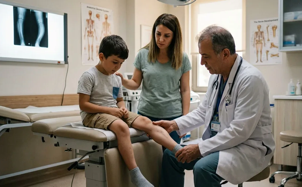
Diagnostic Methods and Clinical Assessment
Diagnosis begins with a thorough physical exam and radiological imaging. The physician assesses deformity severity, joint range of motion, and ligament status. X-rays clearly show bone deficiencies and angular deformities, while MRI provides information about cartilage and soft tissue anomalies.
Accurate measurement of leg length discrepancy is essential for surgical planning.
In advanced cases, 3D modelling via computed tomography (CT) scans allows detailed bone anatomy evaluation. These assessments help define the optimal surgical strategy for lower limb deformities. Diagnosis also involves considering family expectations and the child’s social life, facilitating a personalized treatment roadmap.
Treatment Approach with the Paley Technique
The Paley technique, developed by Dr. Dror Paley, is a comprehensive method used mainly for correcting congenital deformities and limb lengthening. It harnesses the bone’s natural healing capacity to stimulate new bone formation (osteogenesis) while gradually stretching surrounding soft tissues. Compared to conventional methods, it allows correction of more complex deformities and preservation of functional limbs.
This technique is considered a milestone in the field of lower extremity deformity surgery. It relies on 3D deformity analysis and accurate identification of the deformity’s center of rotation (CORA).
Surgical Process and Planning
Preoperative planning is vital for the success of the Paley technique. The surgeon precisely determines the osteotomy site on the bone, type of external fixator device, and mounting angle. Principles of bone lengthening surgery are applied. Special techniques protect nerves and blood vessels during surgery. In fibular hemimelia cases, procedures such as "SUPER hip" and "SUPER knee" are used to stabilize hip and knee joints.
Lengthening and Correction Procedures
Lengthening starts a few days after surgery, usually at a rate of 1 mm per day in gradual steps. External fixators, such as Ilizarov or newer computer-assisted devices, are used. Similar to Ilizarov bone lengthening, new bone forms in the gap created by gradually separating bone ends. Angular deformities are corrected simultaneously.
Recovery and Follow-Up
The treatment does not end with surgery; postoperative care is crucial for sustained success. After lengthening, a “consolidation” phase follows, during which the new bone hardens enough to bear weight. Intensive physiotherapy is required to maintain joint range of motion and increase muscle strength.
Since children continue to grow, regular follow-up until the end of adolescence is necessary. Deformities may recur during growth spurts and new length discrepancies can develop. Long-term cooperation between physician and family is essential. Family compliance significantly influences rehabilitation outcomes.
Risks, Complications and Alternatives
As with any surgery, the Paley technique and deformity surgery carry risks, including pin site infections, joint stiffness, nerve injuries, or delayed bone healing. Conditions necessitating nonunion surgery are rare but possible complications.
Alternatives depend on deformity severity and may include prosthetic use or, in certain cases, amputation. Current practice aims to preserve and reconstruct the limb whenever feasible. The optimal method is chosen after detailed evaluation and shared decision-making with the family.
Information and Disclaimers
Treatment of rare childhood deformities is a highly specialized field requiring extensive experience. This article is intended to inform families about the process. Since each child’s anatomy and deformity severity are unique, treatment plans must be personalized. The most reliable information and treatment options should be obtained through clinical evaluation by an experienced specialist.
This content is provided for general informational purposes only and does not constitute medical advice. It is not intended to replace professional diagnosis, treatment, or guidance. Diagnosis and treatment should only be determined through an in-person consultation with a qualified healthcare provider. Since each patient's clinical condition is unique, surgical or non-surgical interventions may vary accordingly. The information presented is based on current scientific sources and up-to-date medical practices. Do not delay seeking necessary care or professional consultation based on this content.