Bow leg deformity, medically known as "genu varum," is characterized by legs curving outward from the knees. This condition is not just an aesthetic issue but can lead to serious joint load distribution problems if left untreated. Bow leg correction procedures should be carefully planned to improve the patient’s quality of life and to protect joint health in the long term.
Treatment for bow leg correction varies depending on the severity of the deformity, the patient’s age, and bone structure condition. While mild bowing in early childhood is often physiological, persistent or late-onset cases may require intervention. Modern orthopedic approaches offer effective solutions through both surgical and nonsurgical methods.
What Is Bow Leg (Genu Varum)?
Bow leg (genu varum) occurs when the ankles touch while standing but the knees remain apart. This misalignment causes excessive stress on the inner part of the knee (medial compartment), leading to uneven weight distribution. Over time, this imbalance can damage cartilage and cause pain.
Bow Leg Appearance and Clinical Signs
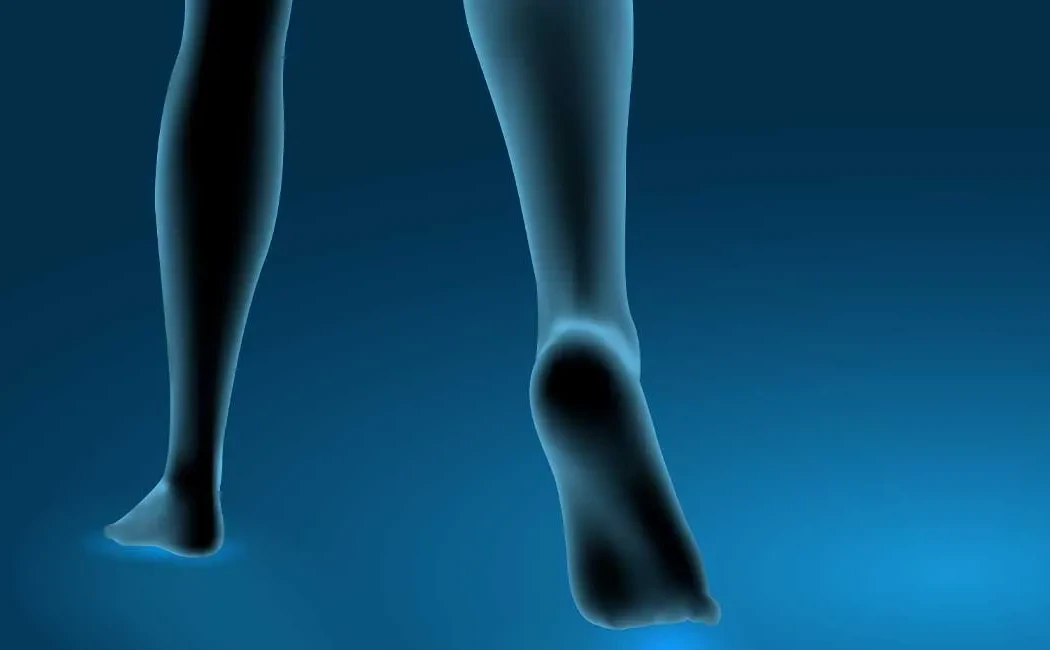
The most noticeable sign is the legs forming a parenthesis shape "( )" when viewed from the front. Patients often report their knees drifting outward or wobbling during walking. Severe cases may present with abnormal gait patterns (such as waddling gait) and rapid fatigue. Clinical evaluation is essential to assess the deformity’s severity.
Who Is Affected?
Bow leg deformity can occur at any age, although causes differ. In infants and toddlers, mild bowing is often physiological and expected to improve over time. However, persistent or newly developed deformities during adolescence or adulthood are considered pathological. In older adults, degenerative joint disease may cause or worsen the deformity.
Causes of Bow Leg Deformity
Accurate identification of the underlying cause is critical for successful bow leg correction. Causes generally include developmental, genetic, or metabolic origins.
Developmental and Genetic Factors
Asymmetric growth of the bone growth plates can lead to curved bone development. Blount’s disease, a significant developmental disorder caused by dysfunction in the inner growth plate of the tibia, causes progressive bowing. Genetic predisposition, skeletal dysplasias, and family history also play roles in deformity formation.
Metabolic and Systemic Diseases
Metabolic conditions affecting bone quality increase deformity risk. Rickets, caused by vitamin D deficiency, is the most common metabolic cause leading to softening and bending of bones in children. In adults, osteomalacia or Paget’s disease can reduce bone strength, predisposing to bowing. Malunited fractures after trauma can also cause mechanical axis deviation.
Diagnosis and Evaluation Methods
A thorough diagnostic process is essential for effective treatment. After taking medical history, the physician performs a detailed physical exam and imaging studies.
Physical Examination and Clinical Assessment
During physical exam, standing and walking postures are observed. The distance between the knees with ankles together (intercondylar distance) is measured. Joint range of motion, ligament stability, and leg length discrepancies are assessed. Gait analysis provides important information about functional impact.
Radiological Imaging
Accurate diagnosis and surgical planning require imaging. In addition to standard X-rays, a full-length lower limb weight-bearing radiograph ("long leg film") is taken from hip to ankle. Mechanical axis deviation (MAD) and joint angles are precisely measured on this film. MRI or CT scans may be requested to evaluate cartilage and soft tissues if needed.
Bow Leg Correction Methods
Treatment depends on the cause, severity of deformity, and patient age. Within the scope of lower limb deformity surgery, the most suitable method is chosen to create a personalized plan.
Non-Surgical Methods
Physiological bow leg cases in early childhood are usually monitored without intervention, expecting spontaneous improvement. Metabolic deformities such as rickets require medical treatment first (vitamin D and calcium supplementation). For mild deformities or patients unsuitable for surgery, special insoles or knee braces can help manage pain and balance joint load.
Surgical Intervention: Osteotomy
In adults and children with progressive deformity, surgery is the most effective bow leg correction method. Osteotomy involves a controlled cut of the bone, usually at the tibia or femur, followed by realignment and fixation with plates, screws, or fixators.
Sometimes deformity correction is combined with limb lengthening procedures when required. Here, bone lengthening surgery techniques can be integrated with osteotomy.
Ilizarov and Limb Reconstruction Techniques
For complex, multi-planar deformities or those accompanied by severe shortening, Ilizarov external fixators or computer-assisted hexapod systems may be preferred. These enable gradual correction of the bone. Due to possible challenges in limb reconstruction surgery, such procedures should be managed by an experienced team.
Post-Operative Recovery and Rehabilitation
The healing process varies depending on the method used. Rehabilitation during bone healing is crucial.
Physical Therapy and Supportive Devices
Early post-operative physical therapy aims to maintain joint mobility and strengthen muscles. Patients may need crutches or a walker for support. Care principles after limb lengthening surgery also apply here; wound care, swelling control, and regular exercises accelerate healing.
Complications and Follow-up
As with any surgery, bow leg correction carries risks such as infection, nerve injury, or non-union. Rarely, treatment for delayed union or malunion may be needed. Surgical approaches for non-union in femoral fractures should be considered if healing is delayed.
Bow Legs in Children and Early Intervention
Bow leg deformities in children can cause parental concern, but distinguishing physiological from pathological bowing is important. Treatment of bow legs (genu varum) in children typically depends on age and growth potential. Early diagnosed Blount’s disease or rickets can often be corrected with growth modulation surgeries such as hemiepiphysiodesis. Pediatric lower limb deformity treatment requires a multidisciplinary approach.
Possible Problems If Left Untreated
Untreated bow leg deformity is not merely cosmetic. Misalignment leads to uneven load distribution on the knee’s inner cartilage, accelerating wear and pain. This results in early onset osteoarthritis (gonarthrosis), meniscus tears, and chronic knee discomfort. In advanced stages, patients may require total knee replacement. Timely correction helps prolong the lifespan of the natural joint.
Medical Disclaimer and Liability Waiver This content is provided for informational purposes only. For diagnosis and treatment, please consult an orthopedic specialist. Each patient's condition is unique and requires personalized evaluation. The information on this site does not replace a professional medical examination or consultation. Do not delay seeking professional care based on the information provided.