Bowleg deformity, medically known as "Genu Varum," is an orthopedic condition characterized by noticeable space between the knees when the ankles are together. This results in an outward curvature of the legs and is commonly identified during childhood. If left untreated, it can lead to both aesthetic concerns and significant functional problems in adulthood.
Bowleg deformity should not be considered merely a cosmetic issue. Misalignment in the legs causes excessive load on the inner part of the knee joint when bearing body weight. Over time, this can lead to cartilage damage, meniscal tears, and early-onset osteoarthritis. Therefore, accurately determining the severity of the deformity and establishing an appropriate treatment plan is critical for long-term joint health preservation.
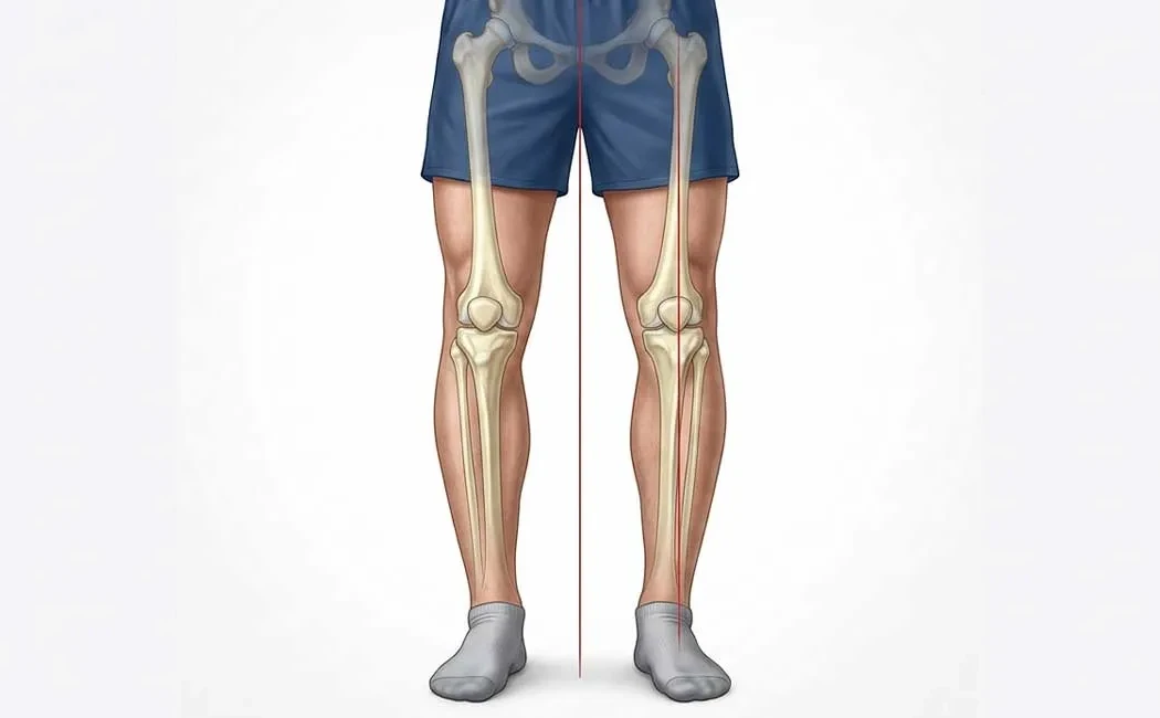
What Is Bowleg Deformity (Genu Varum)?
Genu Varum is an angular deformity resulting from inward deviation of the lower limb’s mechanical axis. In normal leg alignment, the centers of the hip, knee, and ankle lie on the same mechanical axis, whereas in bowleg deformity, the knee joint shifts outward from this axis. This can cause a distinctive walking pattern often referred to as a "cowboy gait."
Physiological (developmental) genu varum is common in infants and toddlers and generally resolves spontaneously by ages 2-3. However, pathological genu varum does not self-correct and tends to progress; it requires medical assessment and intervention.
Causes of Bowleg Deformity
Bowleg deformity can arise from various factors affecting bone development. These causes are broadly categorized as congenital and acquired.
Congenital Causes
In some cases, the deformity is present at birth due to positional constraints in the womb or genetic factors. Skeletal dysplasias, genetic syndromes affecting bone growth, and metabolic bone diseases fall into this category. Early evaluation of congenital deformities and surgical options is advised, as outlined in congenital deformities and surgical treatments. Growth plate disorders such as Blount disease (tibia vara) are important congenital causes of progressive bowleg deformity in childhood.
Acquired Causes
Acquired causes typically develop in childhood or adulthood. The most common is rickets, due to vitamin D deficiency. Trauma affecting growth plates, malunited fractures, bone infections (osteomyelitis), and tumors can also lead to acquired deformities. In adults, advanced knee osteoarthritis with joint space narrowing may cause leg bowing.
Symptoms and Diagnostic Methods
Diagnosis is made through detailed physical examination and imaging studies. While cosmetic concerns are often the main complaint, underlying functional problems are also significant symptoms.
Physical Signs
The most obvious sign is that the knees do not touch when standing, creating a noticeable bow shape to the legs. Other signs include outward knee thrust during walking (lateral thrust), easy fatigue, inner knee pain, and progressively developing limping. Secondary pain may also occur in the ankle and hip joints in some cases.
Imaging and Measurement Techniques
Definitive diagnosis and surgical planning require special full-leg standing X-rays called orthoroentgenograms, showing the entire leg from hip to ankle. Measurements of mechanical axis deviation (MAD) and joint angles are taken.
If necessary, computed tomography (CT) scans assess bone rotational deformities. These measurements are essential for determining the center of rotation of angulation (CORA) and calculating the degree of correction required.
Treatment Options for Bowleg Deformity
Treatment depends on the patient’s age, severity, and underlying cause of the deformity. The primary goal is to correct the mechanical axis to balance load distribution on the knee joint and protect joint health. Complex cases may require lower extremity deformity surgery techniques.
Non-Surgical Methods
Physiological curvature in early childhood often only needs observation. Metabolic causes like rickets require medical treatment to improve bone quality first. In growing children, bowleg deformity treatment may involve custom orthoses and night splints. Additionally, muscle strengthening exercises for lower extremity deformities in children can be beneficial.
Surgical Methods
Progressive deformities, growth plate problems, or adult cases may require surgery. Options include hemiepiphysiodesis (growth modulation) and osteotomy (cutting and realigning bone). In adults and skeletally mature individuals, deformity correction surgery realigns the bone at the correct angle and fixes it with plates, screws, or external fixators.
These surgeries are classified as joint-preserving procedures, which can delay or eliminate the need for joint replacement.
Ilizarov and computer-assisted (hexapod) fixators provide high precision, especially in complex multiplanar deformities.
Postoperative Care and Rehabilitation
Recovery after surgery varies by the method used. Bone healing following osteotomy typically takes 6-8 weeks. During this period, crutches and limited weight-bearing may be necessary. Similar to post-limb lengthening care, routine wound care, pin site maintenance (if fixators are used), and physical therapy are essential after deformity surgery. Rehabilitation aims to maintain joint range of motion and restore muscle strength.
Possible Complications and Risks
As with any surgical procedure, genu varum surgeries carry risks. These include infection, nerve injury, and vascular damage, although rare. One significant complication is delayed or non-union of the bone. In such cases, additional interventions such as non-union surgery may be necessary. Inadequate deformity correction or recurrence over time are also potential risks.
Importance of Early Diagnosis and Intervention
Early diagnosis provides advantages in treatment variety and success rates. Curvatures identified in childhood can often be corrected by less invasive growth modulation techniques, while delayed cases may need more extensive bone surgeries. Timely diagnosis and treatment of deformities in children impact their psychosocial development and physical activity capacity, as noted in early diagnosis and intervention in pediatric deformities. For adults, early intervention prevents irreversible knee joint damage and improves quality of life.
In conclusion, bowleg deformity is a treatable condition. With accurate diagnosis, individualized planning, and expert care, achieving healthy leg alignment and functional anatomy is possible.
This content is provided for general informational purposes only and does not constitute medical advice. It is not a substitute for personal diagnosis, treatment, or professional guidance. Diagnostic and therapeutic decisions should only be made following an in-person consultation with a qualified healthcare provider. Individual patient conditions vary, and therefore, surgical or non-surgical treatments may differ accordingly. The information presented here is based on current scientific knowledge and up-to-date medical practices. Do not delay seeking medical care or consult a healthcare professional for advice specific to your situation.