Genu varum and genu valgum are common deformities encountered in orthopedic and traumatology practice that affect the mechanical alignment of the lower extremities. Commonly known as "O-legs" and "X-legs," these conditions are more than cosmetic concerns; if untreated, they may lead to functional problems threatening joint health. Proper leg alignment is essential for balanced weight distribution across joints and a healthy gait pattern.
This guide explores the causes, symptoms, and current treatment approaches for these lower limb deformities. Particular attention is given to distinguishing between physiological developmental variations in children and pathological cases that require surgical intervention.
What Are Genu Varum and Genu Valgum?
Genu varum and genu valgum refer to deviations from the normal anatomical axis of the knee joint. In a normally aligned leg, the centers of the hip, knee, and ankle lie along the same mechanical axis. Deviations from this axis disrupt load distribution, placing excessive pressure on specific parts of the joint.
Genu Varum (O-Leg): Characterized by knees positioned apart while the ankles touch. Legs bow outward, resulting in increased load on the inner (medial) compartment of the knee.
Genu Valgum (X-Leg): Characterized by knees that touch while the ankles are apart. Legs angle inward, increasing load on the outer (lateral) compartment of the knee.
Causes and Risk Factors
Various factors, from genetic predispositions to metabolic diseases, can contribute to these deformities. It is essential to differentiate developmental temporary bowing from pathological conditions.
Common causes include:
- Physiological Development: Bowing seen at specific ages in children that typically self-corrects.
- Rickets (Vitamin D Deficiency): A metabolic disorder that softens bones, leading to bending.
- Blount’s Disease: A growth disturbance in the upper part of the tibia causing progressive bowing.
- Trauma and Infections: Fractures or osteomyelitis affecting growth plates can result in asymmetric growth.
- Genetic Skeletal Dysplasias: Hereditary disorders affecting the skeletal system.
Symptoms and Clinical Findings
Patients or parents usually notice asymmetry in leg appearance prompting medical evaluation. Alongside deformity, various clinical signs may be present depending on deformity severity.
Common symptoms include:
- Knees hitting each other during walking (genu valgum) or legs splaying outward (genu varum).
- Pain in the knee, hip, or ankle.
- Early fatigue and reduced activity tolerance.
- Advanced cases may involve limping or gait abnormalities.
Diagnostic Process
A thorough diagnostic workup is critical for adequate treatment planning. The process starts with patient history and detailed physical examination. The physician observes the patient standing and walking, assesses limb lengths and joint range of motion.
Radiographic imaging is the gold standard to confirm diagnosis and quantify deformity severity. Long-leg standing X-rays ("orthoroentgenogram") capturing from hip to ankle allow measurement of mechanical axis deviations.
Treatment Methods
Treatment depends on the patient's age, the cause, and deformity severity. The primary goal is to restore mechanical alignment to protect joints and improve function. For detailed treatment options, please refer to our article on Treatment of O-Leg and X-Leg in Children.
Non-Surgical Treatments
In mild deformities and presumed physiological cases, non-surgical treatments may be sufficient.
- Observation: Physiological bowing in growing children is monitored periodically.
- Medical Management: Vitamin D and calcium supplementation for metabolic causes like rickets.
- Orthotics and Bracing: Corrective devices may be employed in specific conditions such as early-stage Blount’s disease.
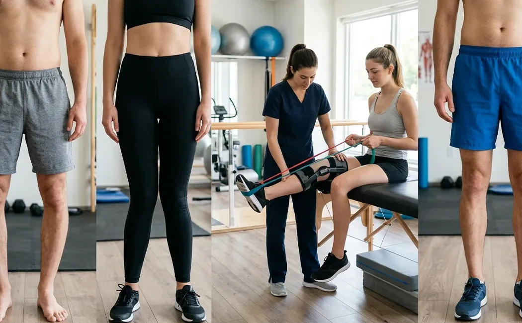
- Physical Therapy: Applied to correct muscle imbalances and maintain joint mobility.
Surgical Treatments
Surgery may be necessary for progressive or severe deformities, and in patients who have reached skeletal maturity. Available surgical options under lower limb deformity correction include:
- Guided Growth Therapy: Small plates or screws are placed on the growth plate on the convex side to slow growth, allowing gradual correction as the other side continues growing.
- Osteotomy: Controlled bone cuts are made to realign the limb, stabilized with plates, screws, or external fixators. Osteotomy is a key joint-preserving procedure.
- Limb Lengthening and Reconstruction: If leg length discrepancy accompanies deformity, methods like the Ilizarov or magnetic nail techniques are used.
- Revision Surgery: Correction of malunions after fractures may be performed using specialized techniques.
Genu Varum and Valgum in Children
Leg bowing is a common finding in children and generally part of normal development. Infants are typically born with slight genu varum (O-legs), which corrects by 18–24 months. Subsequently, genu valgum (X-legs) may develop, usually peaking around ages 3 to 4 and normalizing by ages 7 to 8.
Deformities that fall outside these physiological parameters—such as asymmetry, persistence, or progression—are considered pathological. For more information, visit our Pediatric Lower Limb Deformities page.
Importance of Early Intervention
Early diagnosis significantly influences treatment success in genu varum and valgum cases. Growth plate abnormalities detected early can often be corrected with less invasive surgeries like guided growth, while delayed cases may require more extensive bone surgeries.
Untreated deformities cause irregular load distribution on joint cartilage, leading to premature osteoarthritis. Early intervention in pediatric orthopedics preserves long-term joint health. More details on timing and outcomes are available in our article on Age Factors in Deformity Surgery.
In summary, when managed promptly and appropriately, genu varum and valgum respond well to treatment. Noticing leg deformities in yourself or your child should prompt consultation with an orthopedic specialist to ensure a healthier future.
This content is for general informational purposes only and does not constitute medical advice. It is not intended to replace individual diagnosis, treatment, or professional consultation. Diagnoses and treatment plans should only be made following an in-person examination by a qualified healthcare provider. Since each patient’s clinical condition is unique, surgical or non-surgical interventions may vary accordingly. The information provided is based on current scientific evidence and established medical practices. Do not delay seeking medical care or advice from a healthcare professional based on the information presented herein.